Echocardiogram 2
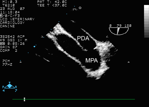
These images are from a one-year-old German Shepherd dog with a large left-to-right shunting patent ductus arteriosus (PDA). The top image shows the PDA from a transesophageal (TEE) view. The transducer is within a flexible endoscopic-like device that has been passed down the esophagus after the dog was anesthetized. The transducer is at the top so the first structure at the top is the aorta (Ao), which lies next to the esophagus. The PDA is immediately below the Ao and communicates with it. The PDA is a long tubular structure that narrows at the end that communicates with the main pulmonary artery (MPA). You can see continuous flow going through the PDA into the MPA. Note that the flow becomes turbulent as it crosses the narrow end of the PDA.
Transesophageal Echocardiograms
Two-dimensional Transesophageal Echocardiogram
Color Flow Doppler Transesophageal Echocardiogram

TEE

From Medicine World.org