Case 1 — Two-Dimensional Echocardiograms
Right parasternal long-axis view
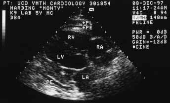
Right parasternal short-axis view

The echocardiograms show a small amount of pericardial effusion (PE) and a moderate amount of pleural effusion (PL). The heart itself appears normal. The hepatic veins were distended on an abdominal ultrasound exam meaning that systemic venous pressure is increased. LV — left ventricular cavity; RV — right ventricular cavity; LA — left atrial cavity; RA — right atrial cavity.