Case 18 — Echocardiograms

Right Parasternal Long-Axis View

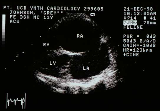
Right parasternal long-axis echocardiogram showing massively enlarged left atrium (LA) and right atrium (RA) with relatively normal-appearing ventricles. RV — right ventricle; LV — left ventricle.
Long-axis view. LA and RA massively enlarged; ventricles relatively normal. RV; LV.

Right Parasternal Long-Axis View (Increased Gain)

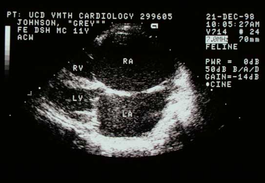
Same view with increased gain showing flocculated densities (spontaneous echocardiographic contrast or 'smoke') swirling within the chambers, especially apparent in the left atrium. This occurs when blood flow velocity slows, allowing red cells to aggregate.
Same view (increased gain). Flocculated densities (spontaneous echocardiographic contrast/“smoke”) swirl within the chambers, especially the LA.

Right Parasternal Short-Axis Basilar View

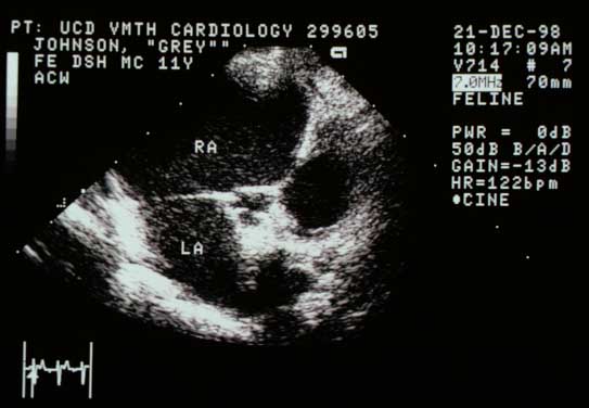
Right parasternal short-axis basilar view with the aorta in the middle, the left atrium (LA) below and to the left, and the right atrium (RA) above. Both atria are massively enlarged.
Short-axis basilar view. Aorta center; LA below left; RA above. Both atria massively enlarged.

Video of an echocardiogram from a different cat with the same disease

Two-dimensional echocardiogram from another cat with a similar disease showing spontaneous echocardiographic contrast


Quiz

1. What primary abnormalities do you see?

2. The spontaneous contrast (“smoke”) occurs when blood flow velocity slows and red cells clump. What complication can occur?