Case 20 — Color Flow Doppler Echocardiograms

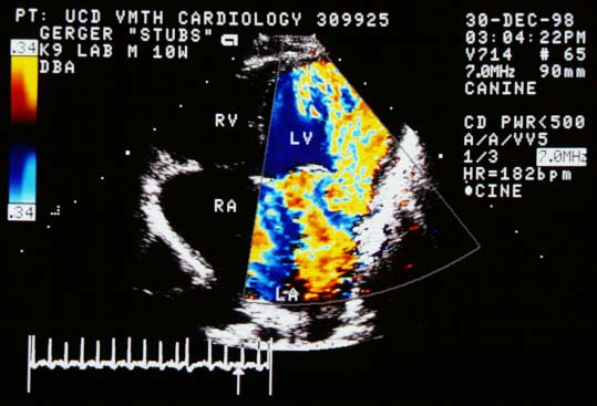
Left apical color flow Doppler in early diastole showing turbulent blood flow projecting from the mitral valve region into the left ventricle.
Left apical view (early diastole). Turbulent flow projects from the mitral valve region into the LV.
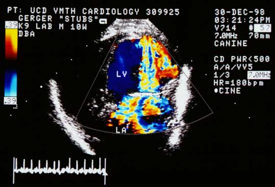
Same left apical view showing flow accelerating toward the mitral valve region and becoming turbulent into the left ventricle in late ventricular diastole.
Same view (late diastole). Flow accelerates toward the mitral valve and becomes turbulent in the LV.
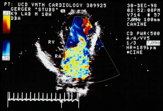
Blood flow appears to flow from the left atrium (LA) through the abnormal left ventricular band to become a turbulent jet in the left ventricle (LV).
Flow from the LA through the abnormal LV band becomes a turbulent jet in the LV.
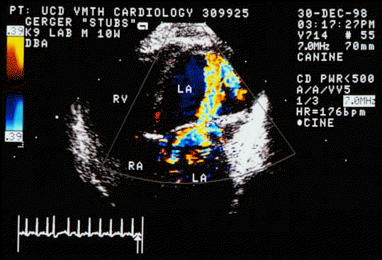
Systolic frame showing blood flowing away from the transducer in the left ventricle (blue), aliasing to gold at the mitral valve (arrowheads), then becoming a turbulent jet in the left atrium — mitral regurgitation.
Systolic frame. LV flow (blue) aliases at the mitral valve (arrowheads) and becomes a turbulent MR jet in the LA.

Color flow Doppler echocardiogram from an animal with the same disease

↑