Case 25 — Radiographs

Lateral

Lateral thoracic radiograph showing an enlarged cardiac silhouette with lucent structures (gas-filled gut loops) within it, consistent with peritoneopericardial diaphragmatic hernia.
Lateral view.

DV

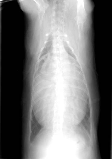
Dorsoventral thoracic radiograph showing an enlarged cardiac silhouette with heterogeneous density including gas-filled structures.
Dorsoventral view.

Quiz

1. What are the lucent structures within the cardiac silhouette?

2. These radiographs are highly suggestive of what abnormality?