Tiffany was diagnosed with mandibular micrognathia with resulting palatal defect from the left mandibular canine at the VMTH on her initial visit. Her owners were given the treatment options of orthodontic appliance or crown amputation with partial pulpectomy. The latter option was strongly advised and chosen by the owners. They returned several days later for the dental procedure. Transesophageal echocardiography was performed during the same anesthetic period.
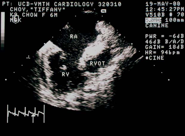
Transesophageal Echocardiography
TEE showing a ridge of tissue between the right ventricle (RV) and the right ventricular outflow tract (RVOT). The right atrium (RA) is severely enlarged. Spontaneous contrast is visible in the RA during diastole due to sluggish blood flow.
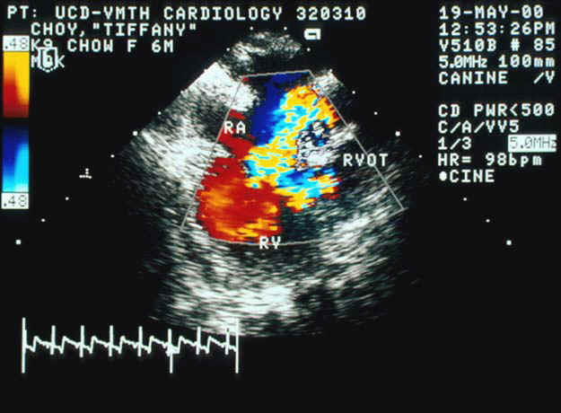
Color Flow Doppler TEE
Color flow Doppler TEE demonstrates turbulent flow from the right ventricle (RV) into the right atrium (RA) due to tricuspid regurgitation and into the right ventricular outflow tract (RVOT) due to the obstruction. CW Doppler again documented peak systolic velocity across the tricuspid valve of approximately 5 m/s.