Case 28 — Ultrasound Exams
Echocardiography
The echocardiogram was normal.
Ultrasound Examination of the Lesion
This is a video clip of color flow Doppler showing the blood flow in the lesion.
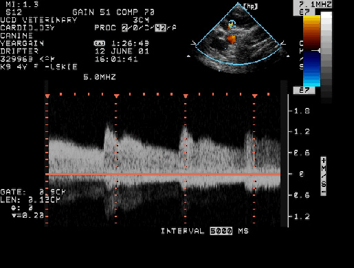
The picture below shows blood flow velocity using pulsed wave Doppler echocardiography. Note that the flow is continuous.