Case 29 — Two-Dimensional Echocardiograms

Right parasternal long-axis view of four chambers
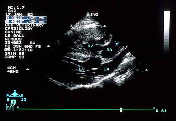
Right parasternal long-axis four-chamber echocardiogram showing right ventricular free wall thickness of 6 mm and left ventricular free wall thickness of 4 mm.
Note that the right ventricular free wall is 6 mm (0.6 cm) thick whereas the left ventricular free wall is 4 mm thick. RV – right ventricular cavity; LV – left ventricular cavity; RA – right atrial cavity; LA – left atrial cavity.

Right parasternal long-axis view of four chambers and aorta
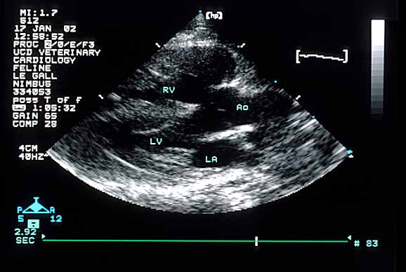
Right parasternal long-axis view showing four chambers and aorta.
RV – right ventricular cavity; LV – left ventricular cavity; Ao – aorta; LA – left atrial cavity.

Right parasternal cranial long-axis view of ventricles and aorta
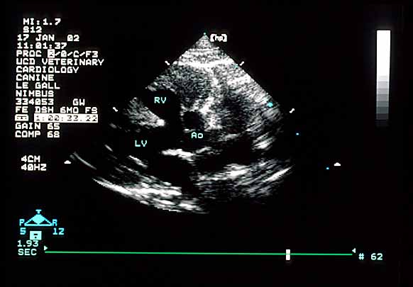
Right parasternal cranial long-axis view showing ventricles and aorta.
RV – right ventricular cavity; LV – left ventricular cavity; Ao – aorta.

Left apical four-chamber view

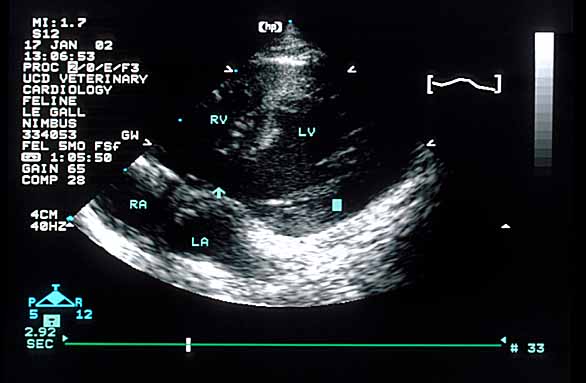
Left apical four-chamber echocardiogram with arrow pointing to ventricular septal defect.
The arrow points to one of the primary abnormalities that makes up this cat’s congenital heart disease. RV – right ventricular cavity; LV – left ventricular cavity; RA – right atrial cavity; LA – left atrial cavity.

Right parasternal cranial short-axis basilar view
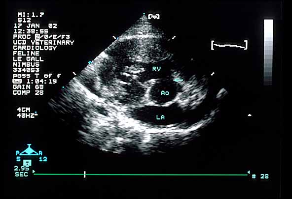
Right parasternal cranial short-axis basilar view with arrow pointing to ventricular septal defect.
The arrow again points to the same abnormality seen in the echocardiogram immediately above this one. Although it looks like the abnormality is between the aorta and right ventricle, it is high on the septum. RV – right ventricular cavity; Ao – aorta; LA – left atrial cavity.

Right parasternal view of the right ventricle and main pulmonary artery
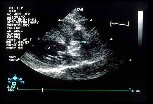
Echocardiogram showing markedly malpositioned pulmonic valve.
This view shows a markedly malpositioned pulmonic valve. The valve is difficult to visualize but sits between the right ventricle (RV) and main pulmonary artery (MPA) at about 2 o’clock, instead of the normal 3 to 4 o’clock position.

Quiz

1. What abnormality(ies) have you identified so far?


↑