Case 31 — Echocardiograms

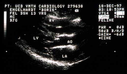
Right parasternal long-axis view

Right parasternal long-axis echocardiogram.
LV – left ventricle; RV – right ventricle; AO – aorta; LA – left atrium

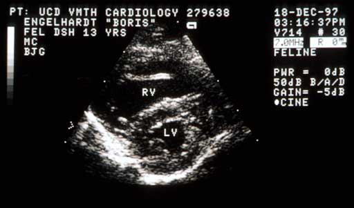
Right parasternal short axis view – systole

Right parasternal short-axis echocardiogram during systole.
RV – right ventricle; LV – left ventricle

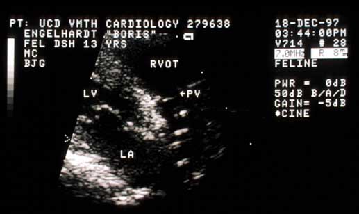
Right parasternal view

Right parasternal echocardiogram showing outflow tract and pulmonic valve.
LV – left ventricle; LA – left atrium; RVOT – right ventricular outflow tract; PV – pulmonic valve

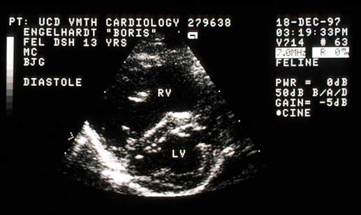
Right parasternal short-axis view – diastole

Right parasternal short-axis echocardiogram during diastole showing septal flattening.
LV – left ventricle; RV – right ventricle

Quiz

1. In the last echocardiogram on this page, the interventricular septum is flattened during diastole. What does this tell you?

2. In the second and fourth echocardiograms on this page you can see that the right ventricular chamber is larger than normal and that the right ventricular free wall is as thick as the left ventricular free wall (i.e., it is too thick). What is this characteristic of?