Case 35 — Pacemaker Implantation
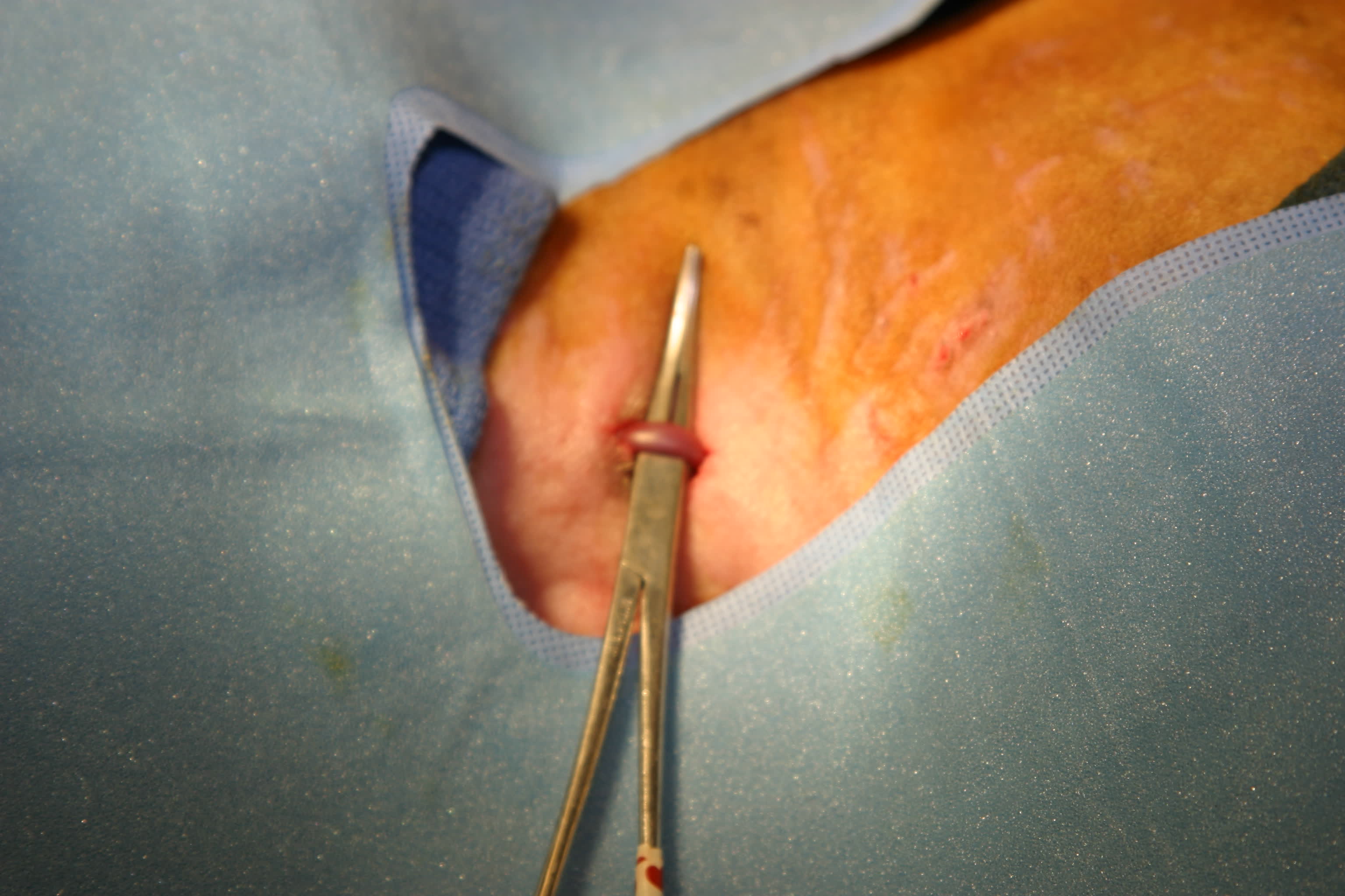
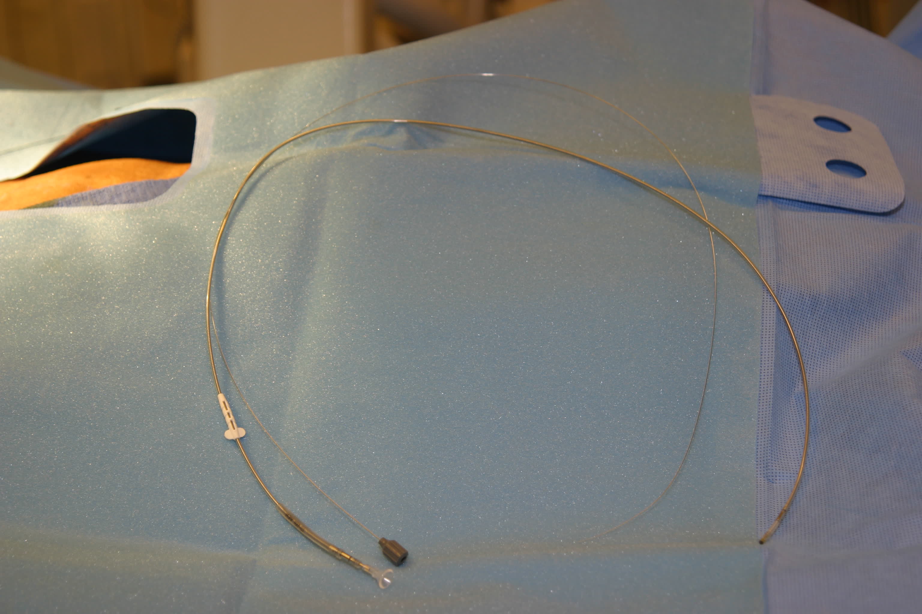
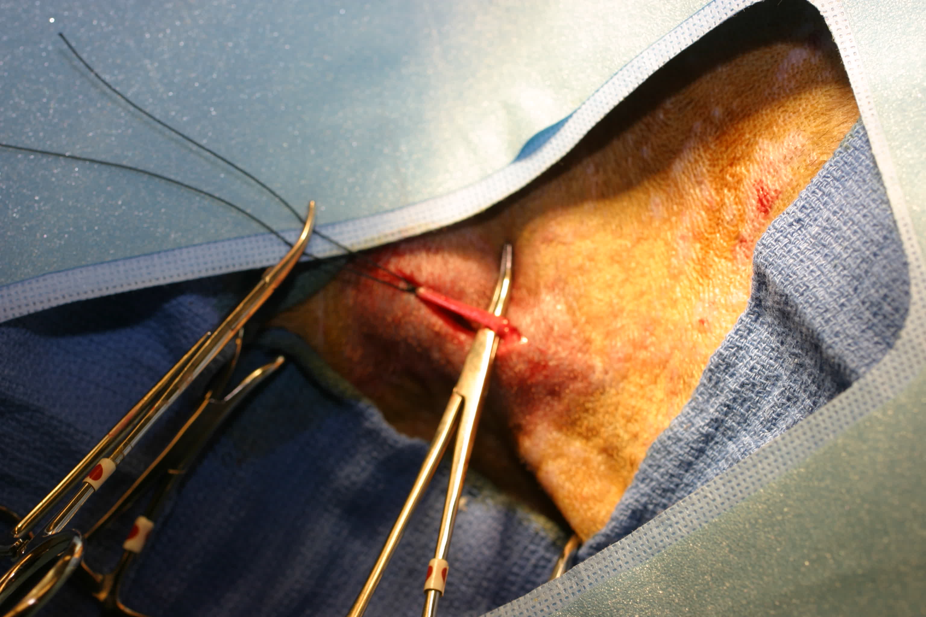
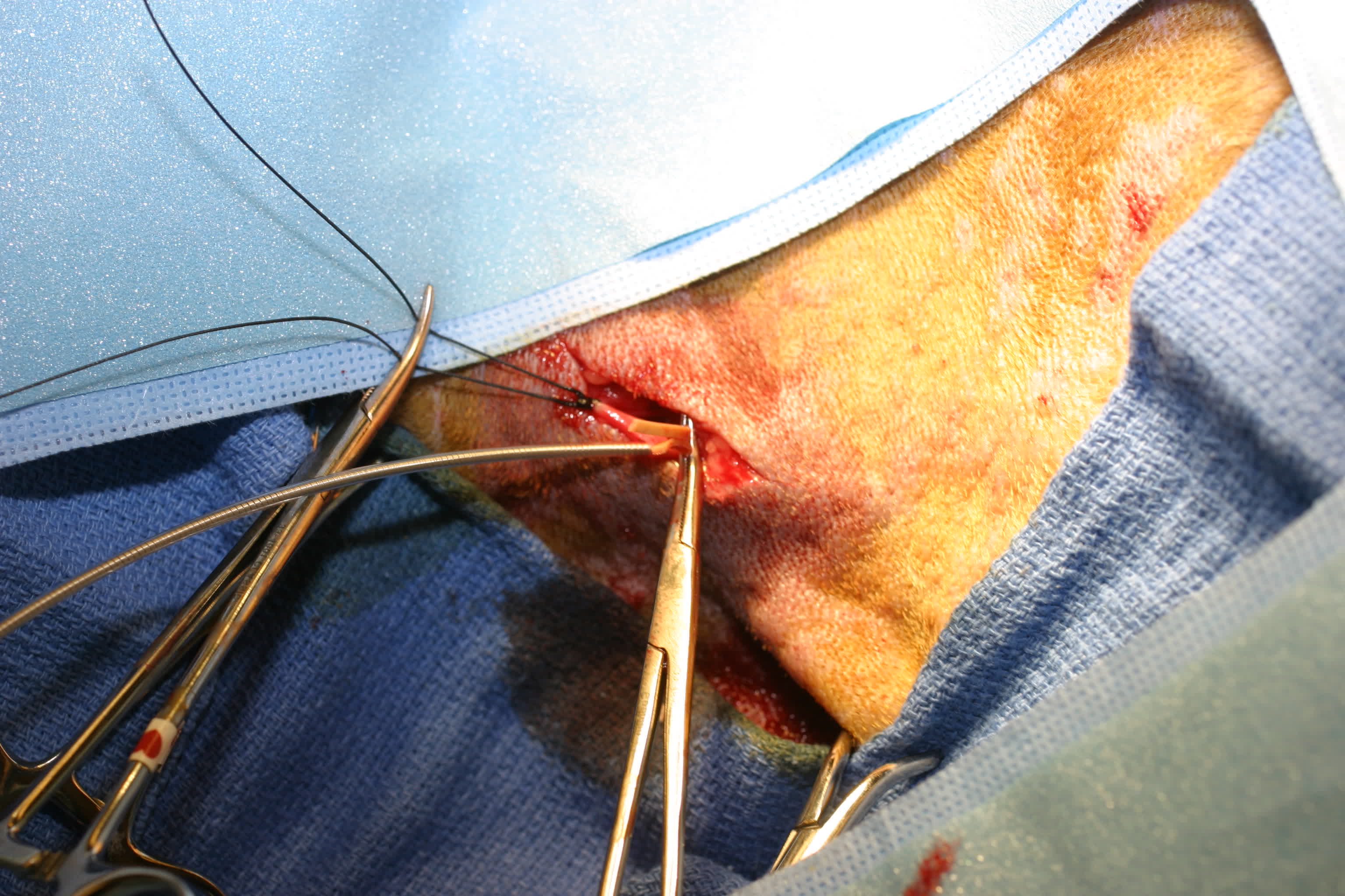
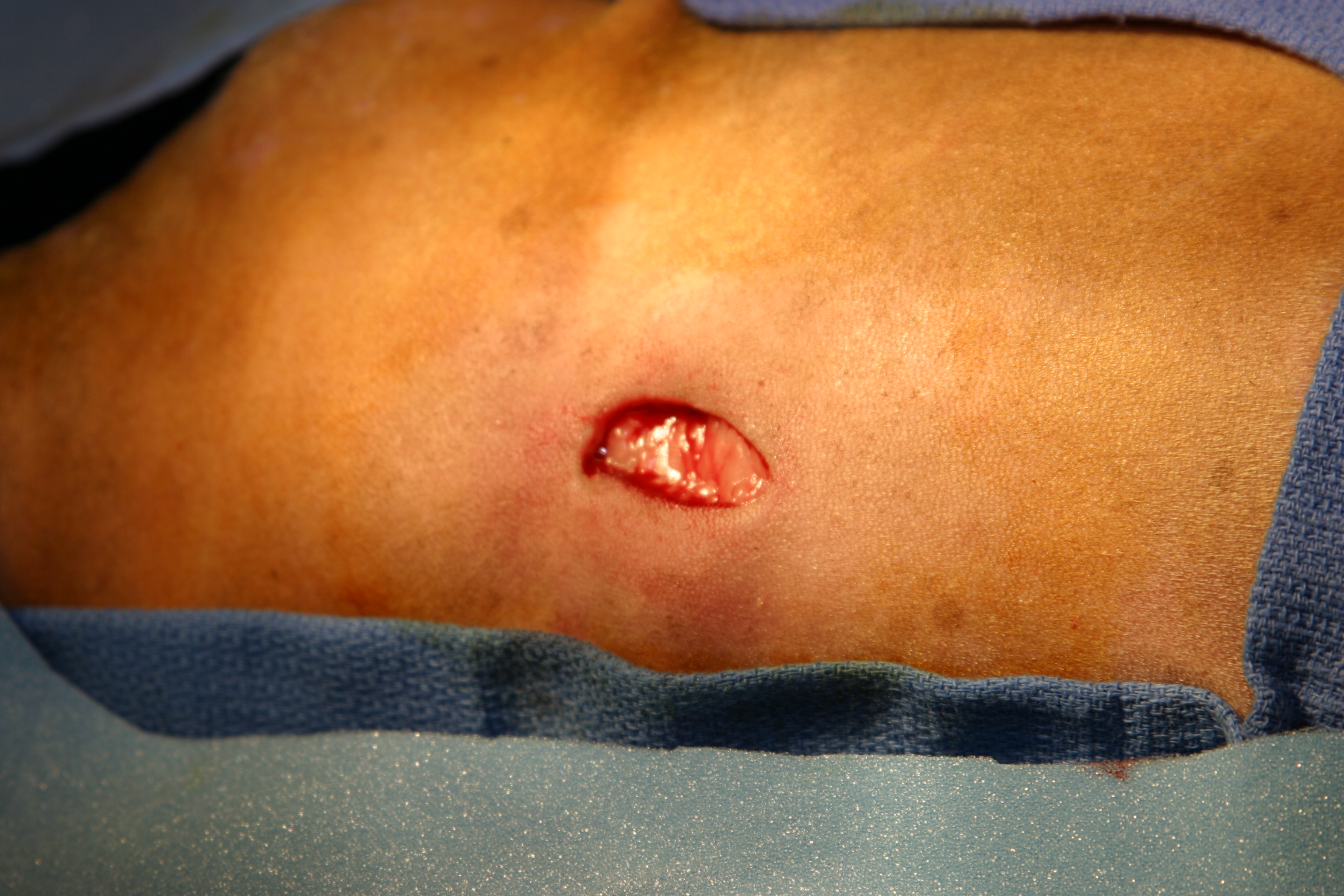
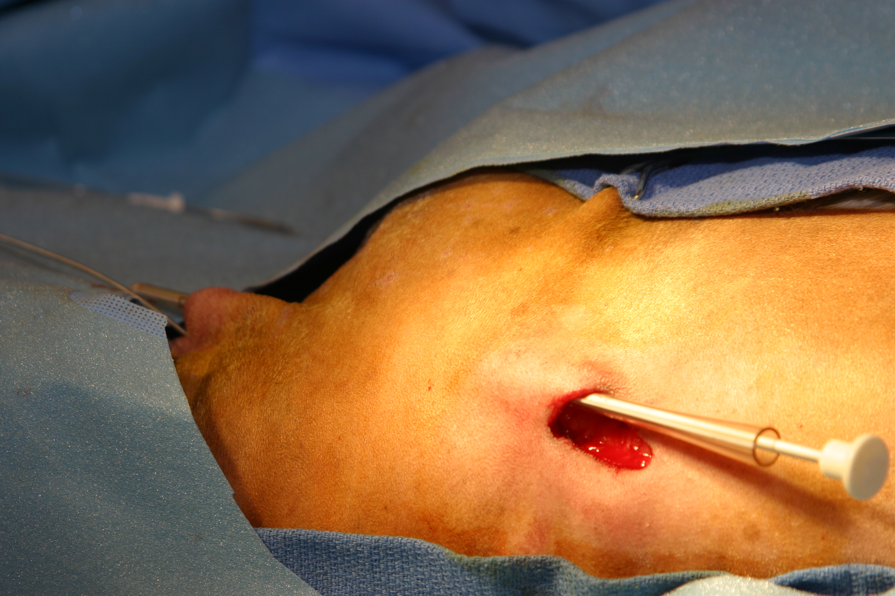
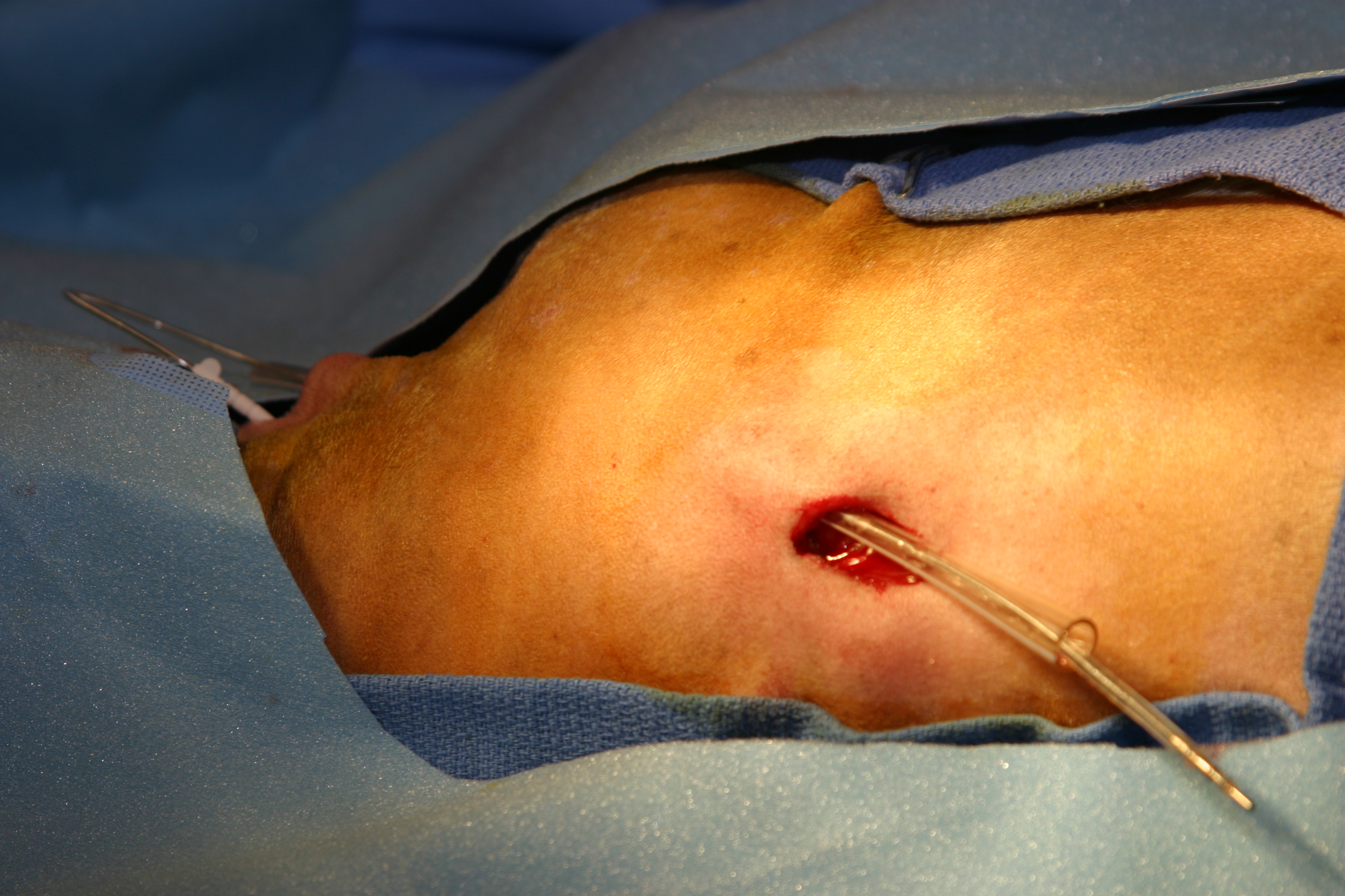
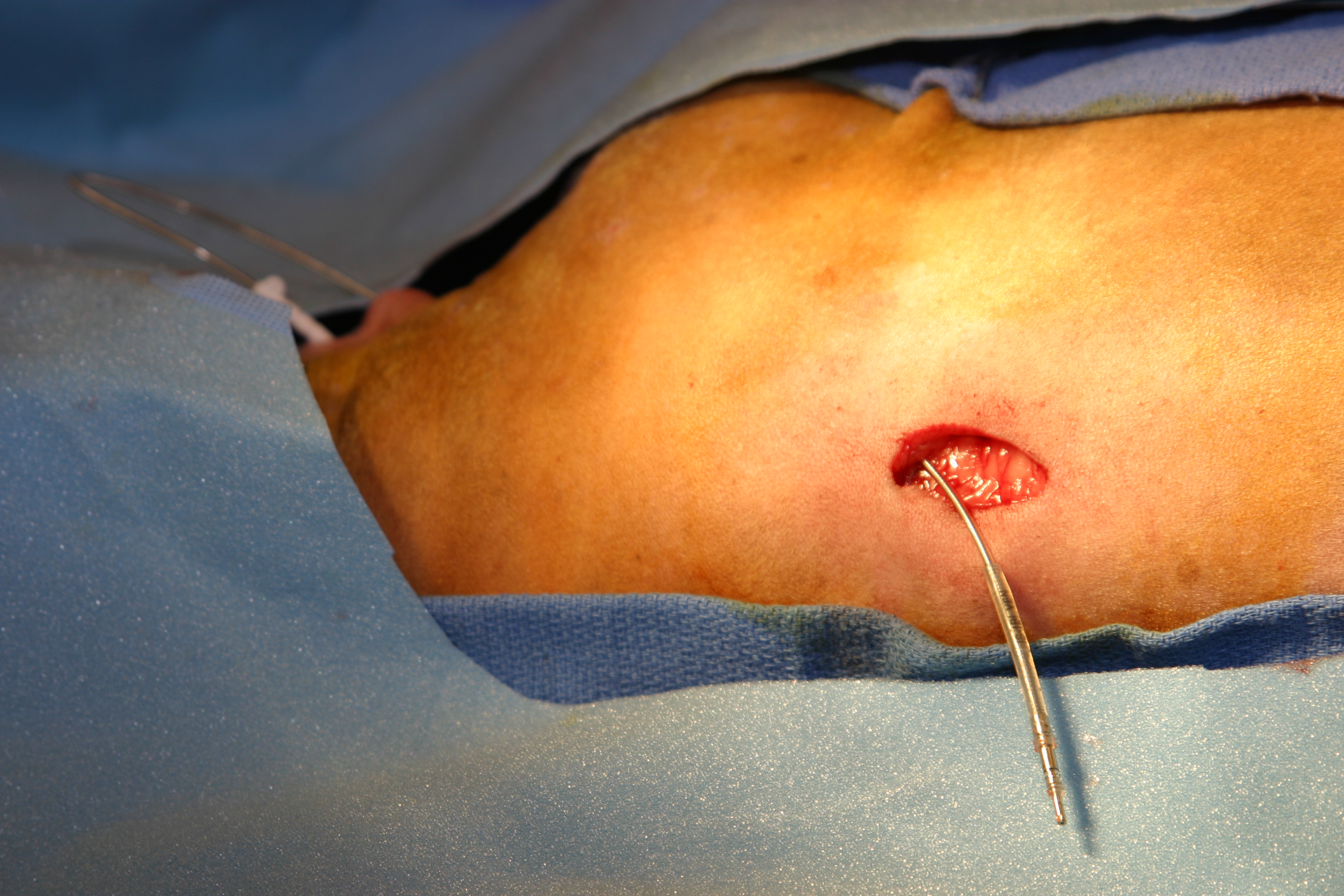
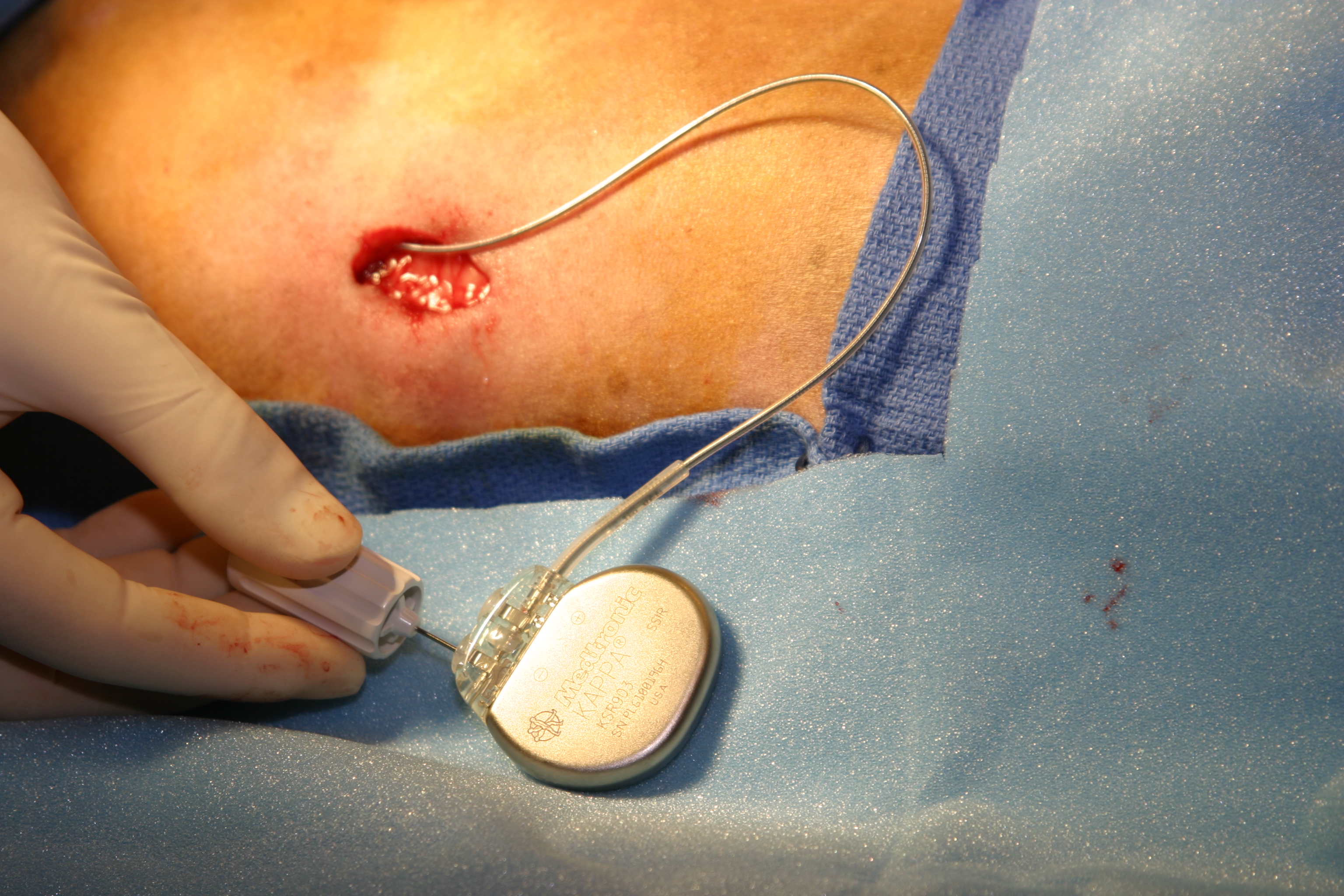
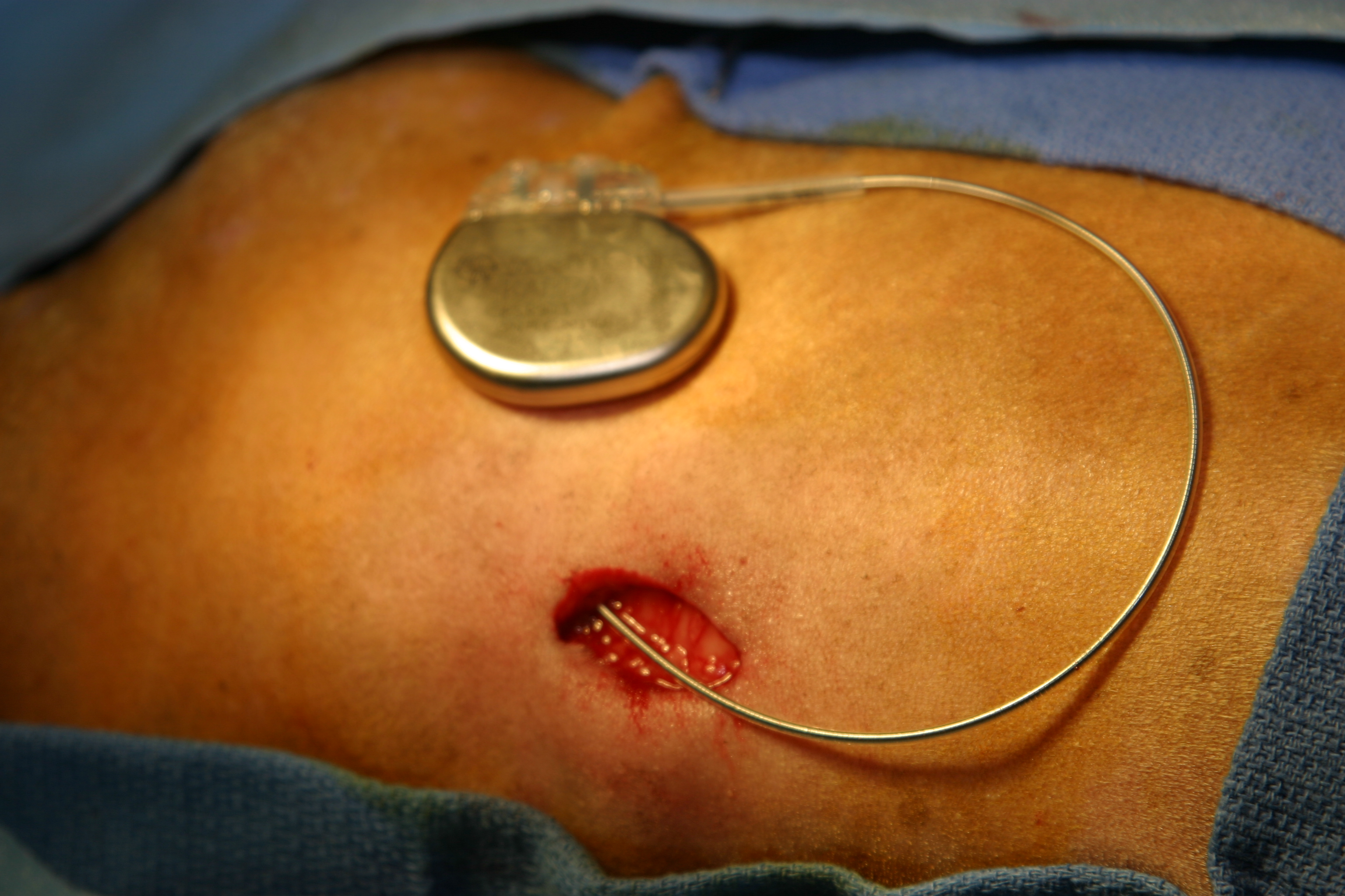
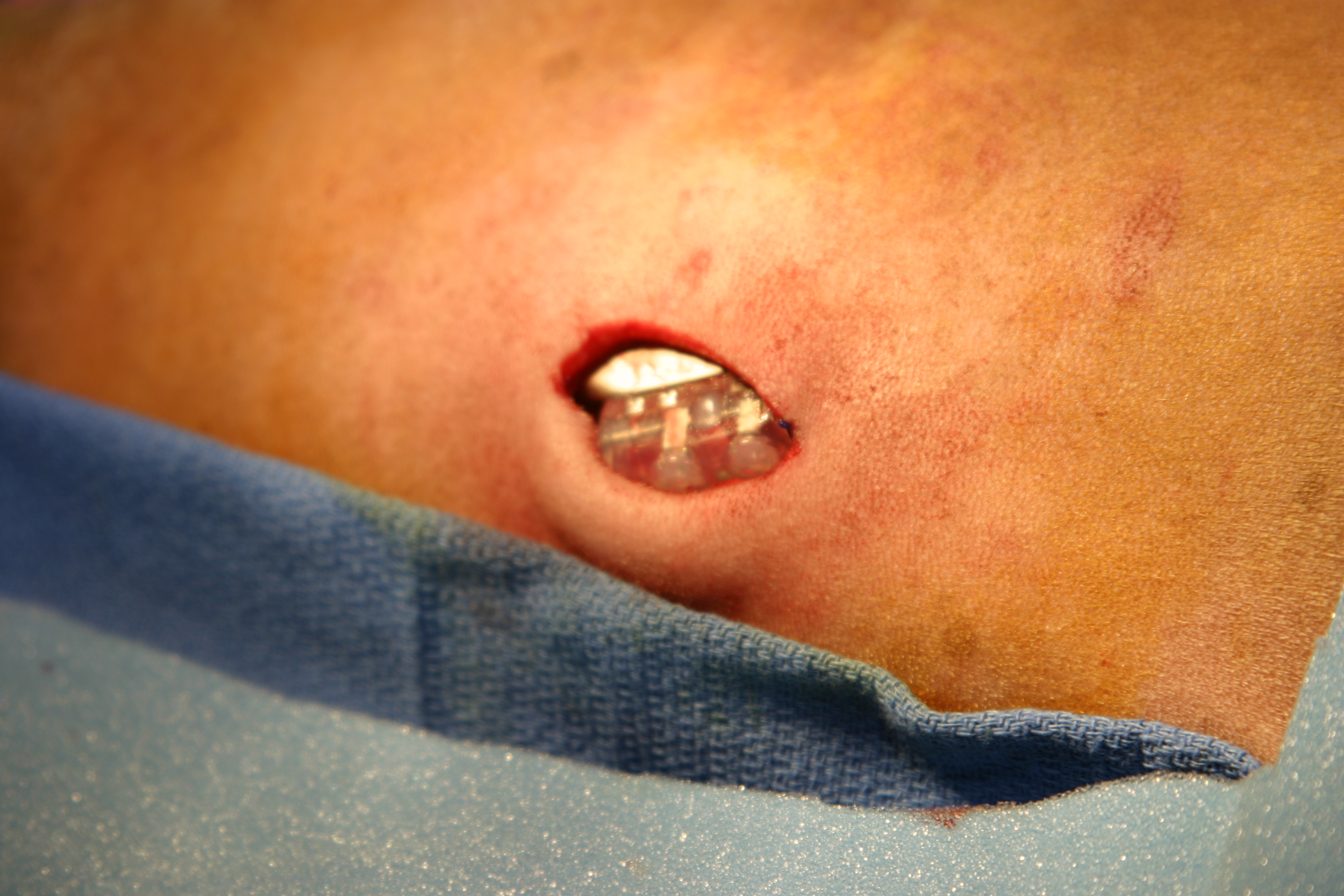
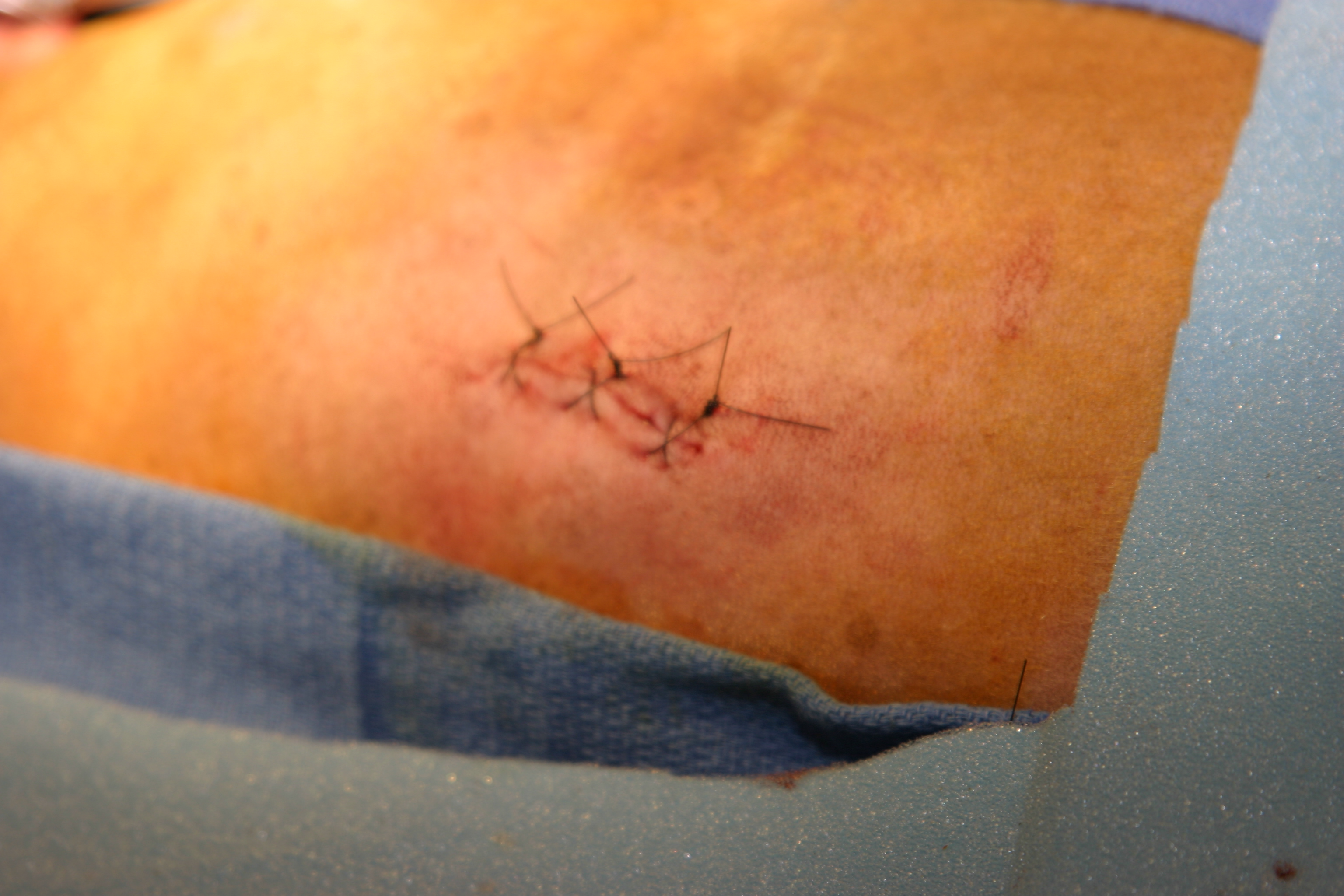
This sequence of images shows the jugular vein being surgically isolated and then a pocket made over the lateral thorax to accommodate the generator. A plastic tube with a trocar is then fed subcutaneously from the pocket on the lateral thorax to the jugular vein incision site. The trocar is removed and the pacing lead fed through it. The pacing lead is already placed with the tip in the right ventricular apex. The generator is then connected to the lead and pacing commences. The incisions are closed. The video shows the pacing system in place under fluoroscopy.