Case 10 — M-Mode Echocardiography
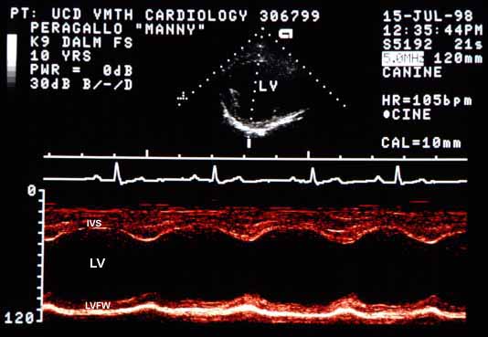
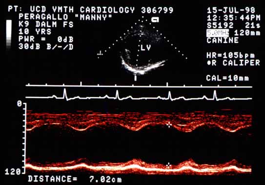

The left ventricular end-diastolic diameter (EDD) is markedly enlarged at 7 cm (normal = 4.7 cm). The end-systolic diameter (ESD) is moderately enlarged at 5 cm (normal = 3.2 cm). The shortening fraction is 29%, within normal range (25–40%). However, myocardial contractility is at least moderately decreased, as evidenced by the moderate increase in end-systolic diameter (the shortening fraction is within normal range only because the end-diastolic diameter is larger than normal). The interventricular septum (IVS) motion is increased (hyperdynamic) while left ventricular free wall (LVFW) motion is reduced. Wall thicknesses are normal (~1 cm), indicating eccentric (volume overload) hypertrophy is present.
The E-point to septal separation (EPSS) was 16 mm, at least moderately increased from the normal of less than 6 mm. The left atrial diameter on a short-axis basilar view was 48 mm compared to an aortic diameter of 20 mm, for a ratio of 2.4 (normal <1.5).