Case 2 — Two-Dimensional Echocardiograms


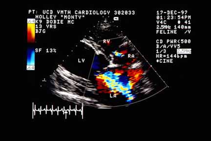
These two-dimensional echocardiograms show the enlarged diastolic diameter of the left ventricle (note that the cursor on the ECG on the two upper pictures is on the QRS complex documenting the frames were taken at end-diastole) and an enlarged left atrium. Note that the left atrium is similarly enlarged to the left ventricle. Dogs with primary mitral regurgitation (usually myxomatous mitral valve disease) generally have a left atrium that is larger than the left ventricle. The right side of the heart is also enlarged but not to the same degree as the left heart. The third picture is taken in systole (cursor between the QRS complex and the T wave) and shows a high-velocity, turbulent jet originating at the mitral valve and extending into the left atrium. Note that the jet stays in the center of the left atrium.