Case 2 — Radiographs

Lateral view

Lateral thoracic radiograph showing moderate to severe cardiomegaly with dorsal displacement of the trachea and elevation of the left mainstem bronchus above the right, consistent with left atrial enlargement

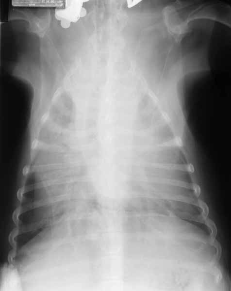
DV view

Dorsoventral thoracic radiograph in which the cardiac silhouette cannot be clearly discerned due to a small amount of pleural effusion obscuring the cardiac borders

Quiz

1. How would you describe the cardiac silhouette on the lateral view?

2. What is the most likely reason the cardiac silhouette cannot be discerned on the DV view?

3. The left mainstem bronchus is elevated slightly above the right mainstem bronchus on the lateral view. This is indicative of: