Case 21 — Echocardiograms
Right Parasternal Short-Axis View of the Left Ventricle

Video of a right parasternal cross-sectional echocardiogram from a different cat with the same cardiac disease
Right Parasternal Short-Axis View of the Left Atrium
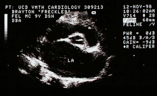
Right Parasternal Long-Axis View of LV and LA
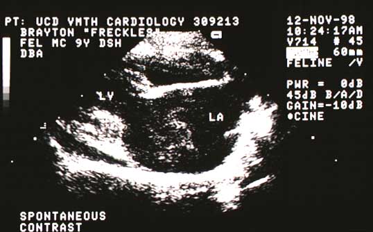
Oblique View of the Left Atrium
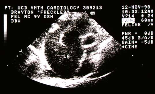
Video of an echocardiogram from a different cat with the same abnormality
Spontaneous contrast in the body of the left atrium and a thrombus in the left auricle (top of the video)