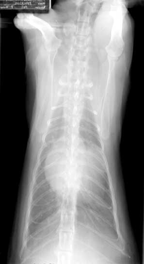
DV radiograph. Enlarged cardiac silhouette with the left auricle touching the left chest wall.
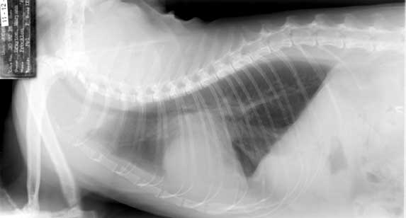
Right Lateral
Right lateral radiograph. Pleural effusion below the last thoracic vertebrae. The left auricle appears as a denser region within the cardiac silhouette.