Skip to main content
SACM
Home
Cases
ECG Atlas
Case 22 — Two-Dimensional Echocardiograms
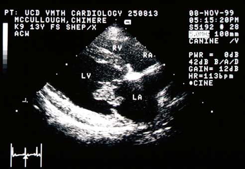
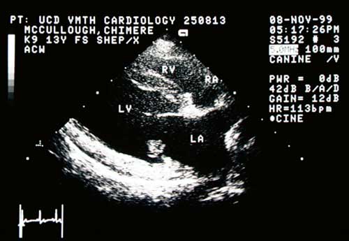
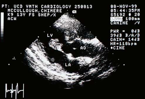
Right Parasternal Long-Axis View
Right parasternal long-axis view.
Right parasternal long-axis view.
Right parasternal long-axis view.
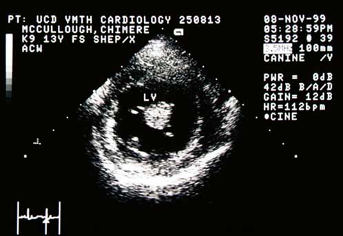
Right Parasternal Short-Axis View
Right parasternal short-axis view.
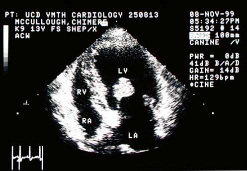
Left Apical Four-Chamber View (Diastole)
Left apical four-chamber view (diastole).
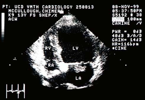
Left Apical Four-Chamber View (Systole)
Left apical four-chamber view (systole).
Video of an echocardiogram from the right parasternal long-axis four-chamber view from a different dog with the same abnormality
Your browser does not support the video tag.
Quiz
1. How would you characterize the abnormality on the septal (anterior) leaflet of the mitral valve?
Gnarley
Cool
Vegetative
Subtle
Check Answer
← Radiographs
Next: Color Flow →
↑