Case 23 — Two-Dimensional Echocardiograms

Right Parasternal Long-Axis (Systole)

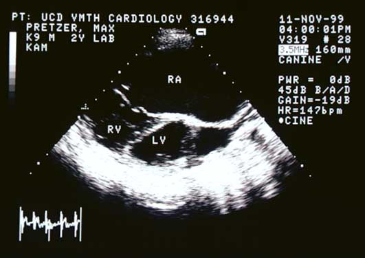
Right parasternal long-axis echocardiogram in systole showing massive right atrial enlargement and dysplastic tricuspid valve.
Right parasternal long-axis view (systole).
Second right parasternal long-axis echocardiogram in systole.
Right parasternal long-axis view (systole).

Left Apical Four-Chamber (Systole)

Left apical four-chamber echocardiogram in systole showing massive right atrial enlargement with the right atrium larger than the right ventricle.
Left apical four-chamber view (systole).

Right Cranial Parasternal (Systole)

Right cranial parasternal echocardiogram in systole.
Right cranial parasternal view (systole).

Video of an echocardiogram (left apical 4-chamber view) from a different dog with the same abnormality


Quiz

1. The right atrium is massively enlarged and is larger than the right ventricle. These findings are strongly suggestive of what functional abnormality?

2. The left heart is smaller than normal. What is the most likely reason for this?