Case 23 — Color Flow Doppler

Left Apical Four-Chamber View (Systole)

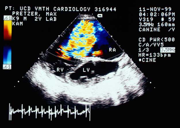
Color flow Doppler in the left apical four-chamber view during systole showing a large jet of tricuspid regurgitation filling the massively enlarged right atrium.
Left apical four-chamber view (systole).

Right Parasternal Long-Axis View (Systole)

Color flow Doppler in the right parasternal long-axis view during systole showing severe tricuspid regurgitation.
Right parasternal long-axis view (systole).

Video of a color flow Doppler echocardiogram from a different dog with the same abnormality


Quiz

1. The color flow Doppler images are diagnostic of what abnormality?

2. How would you grade the severity of this abnormality?