Case 24 — Surgery
The owners were informed of the aggressive nature of a hemangiosarcoma and of the guarded/grave prognosis for Nitro. Treatment options were discussed and the owners were interested in pursuing surgical removal and biopsy of the mass with the option of euthanasia at the time of surgery if metastatic lesions were identified. At this point, no metastatic disease had been found.
Preoperative blood work revealed a mild anemia (PCV = 32%) that was normochromic and normocytic. The rest of the blood work and serum chemistries were unremarkable.
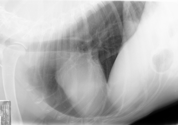
Nitro was anesthetized and a right lateral thoracotomy performed.
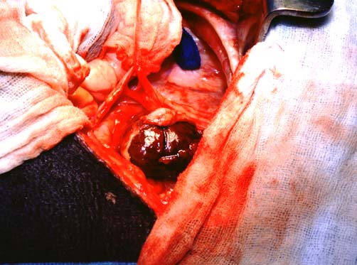
Remarkably, there was no evidence of metastatic disease in any area of the thorax. Consequently, the right auricle was isolated and removed along with the mass and the thorax closed.
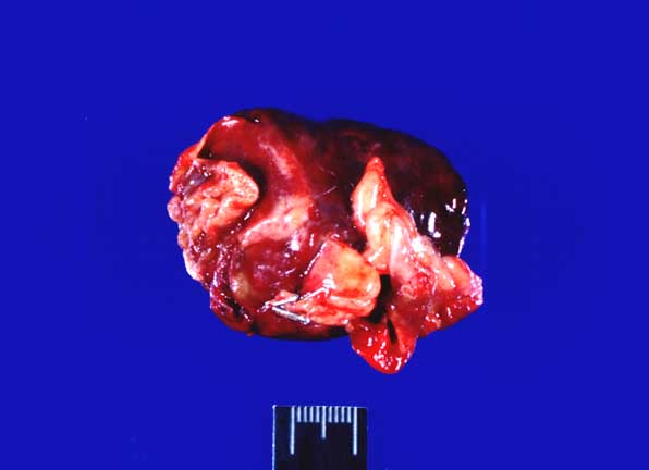
Nitro was taken to the ICU to recover. Histological examination of the mass revealed it was a hemangiosarcoma. The owners were informed that his prognosis was still poor with a median survival of 4 months. Chemotherapy was discussed and offered but the owners declined. Consequently, the only medication he went home on was doxycycline in the hope that it might act as an antiangiogenic agent.