Case 26 — Another Canine Case of Double Chambered Right Ventricle

This is a two-year-old Fox Terrier that presented with no clinical signs referable to heart disease. This dog did not have tricuspid valve dysplasia and so appeared to be having a natural history similar to uncomplicated severe pulmonic stenosis where right heart failure is rare.

Two-Dimensional Echocardiogram (Right Parasternal View)
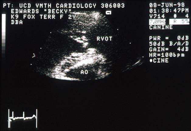
Right parasternal echocardiogram of a Fox Terrier showing a fibrous lesion between the body of the right ventricle and the right ventricular outflow tract, similar to Tiffany's but more fibrous in appearance.
Right parasternal view. This lesion is in the same location as Tiffany’s (between the body of the right ventricle and the RVOT) but appears more fibrous.

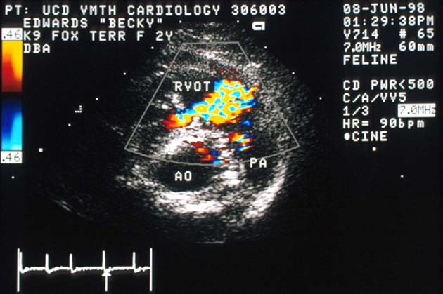
Color Flow Doppler Echocardiogram

Color flow Doppler echocardiogram showing turbulent flow through the obstruction into the right ventricular outflow tract. PA = main pulmonary artery; AO = aorta.
Color flow Doppler shows turbulent flow through the obstruction into the RVOT. PA = main pulmonary artery; AO = aorta.

Continuous Wave Doppler Echocardiogram
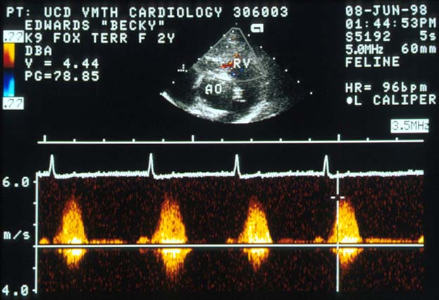
Continuous wave Doppler echocardiogram showing underestimated velocity and pressure gradient due to non-parallel beam alignment, but still placing the lesion in the severe category.
The turbulent flow is not parallel with the continuous wave Doppler beam and so the velocity and pressure gradient are underestimated. Even so, the pressure gradient places the lesion in the severe category.