Case 27 — Radiographs

Lateral

Lateral thoracic radiograph showing a prominent main pulmonary artery and moderate interstitial pattern in the caudal lung lobes.
Lateral view.

Ventro-Dorsal (VD)

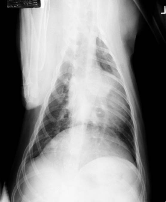
Ventrodorsal thoracic radiograph showing a prominent main pulmonary artery bulge and an enlarged right caudal lobar pulmonary artery.
Ventrodorsal view.

Quiz

1. There is one great vessel that is prominent on both radiographic views. Which one is it?

2. How would you describe this dog’s caudal lobar pulmonary arteries on the VD view?

3. What diagnostic test(s) would you do next given this dog’s history of hemoptysis and the appearance of these radiographs?