Case 33 — Two-Dimensional Echocardiograms

Right parasternal long-axis view

Right parasternal long-axis echocardiogram

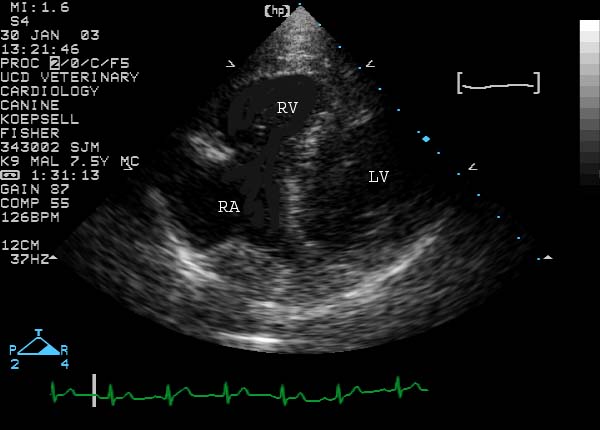
Left apical view

Left apical echocardiogram showing large RV and RA

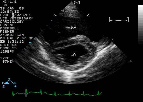
Right parasternal cross-sectional view (diastole)

Cross-sectional echocardiogram in diastole showing RV enlargement and septal flattening

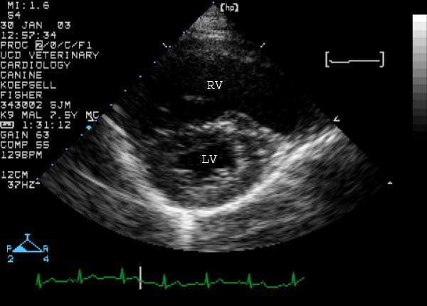
Right parasternal cross-sectional view (systole)

Cross-sectional echocardiogram in systole showing septal flattening

Right parasternal cross-sectional view from a different dog with the same hemodynamic abnormality


Quiz

1. How would you characterize the changes in the right ventricle?

2. What pathophysiologic process would best explain the changes in the right ventricle?

3. What specific disease process would best explain the changes in the right ventricle?

4. The left ventricle is normally round in diastole and in systole. The interventricular septum in this dog is flattened in diastole. Of what significance is this?

5. The interventricular septum is also flattened in systole. Of what significance is this?


↑