Case 37

Two-dimensional echocardiograms

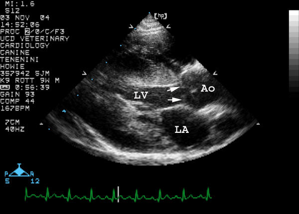
Right parasternal long-axis view
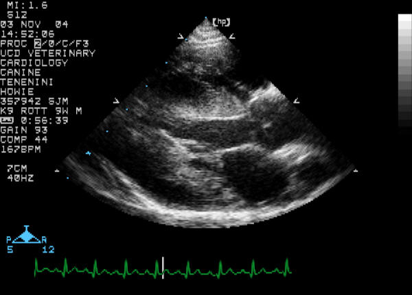
Right parasternal long-axis viewRight parasternal long-axis view labeled

Arrows - Primary lesion


Right parasternal long-axis view video clip

Right parasternal long-axis view of the LVOT and aorta from a different dog with the same lesion


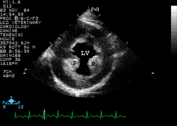
Right parasternal short-axis view of the left ventricle (LV)
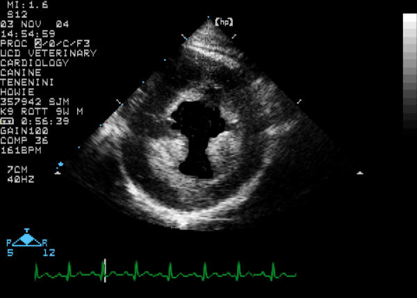
Right parasternal short-axis view of LVRight parasternal short-axis view of LV labeled

LV - left ventricle; P - Papillary muscle

Right parasternal short-axis view of the aorta (Ao) and left atrium (LA)

Right parasternal short-axis view of Ao and LARight parasternal short-axis view of Ao and LA labeled


Quiz

1. The arrows in the top right picture are pointing at what lesion?

2. What would be the best term to call the echocardiographic appearance of the papillary muscles and subendocardium on the short-axis view of the left ventricle?

3. This particular appearance is most likely due to:

4. How would you characterize the size of the left atrium in the short-axis view?