Case 37

Continuous wave Doppler echocardiogram

From a left apical view, a CW Doppler cursor was placed across the left ventricular outflow tract (LVOT) and aorta and this recording of peak blood flow velocity was made. The peak velocity was measured (green box in upper right corner).
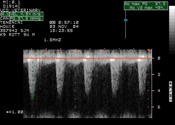
Continuous wave Doppler echocardiogram


Quiz

1. The ultrasound machine has converted the peak velocity of the jet through the lesion (4.94 m/s) to a pressure gradient across the lesion to be 97.6 mmHg. What formula did it use?

2. The velocity and pressure gradient mean the lesion in this dog is: