Case 5 — Two-Dimensional Echocardiogram / Color Flow Doppler
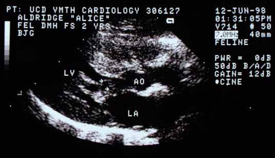
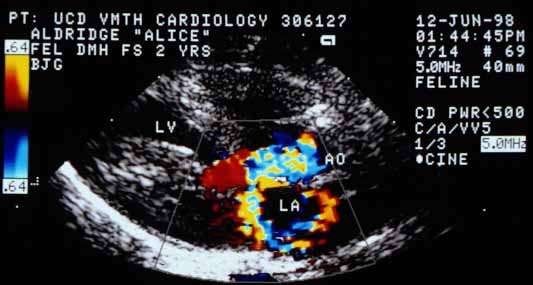
When the color flow Doppler is used to look at blood flow in the same orientation as the view above, two high-velocity jets can be identified originating from the same site in the left ventricle (LV) during systole. One jet projects into the aorta (AO) and the other projects into (and curls around within) the left atrium (LA). Both originate from the site of obstruction seen in the echocardiogram above.
Video clips of echocardiograms from another cat with the same disease
Within this continuous loop of one cardiac cycle you can see the septal (anterior) mitral valve leaflet and chordal structures appear in the left ventricular outflow tract during systole. A portion of the cranial papillary muscle can be seen pulling chordae tendineae into the outflow tract.
Two turbulent jets can be seen — one projecting back into the left atrium and the other going out into the aorta. Both jets originate from the region in the outflow tract where the chordal structures and anterior mitral valve leaflet impinge on the outflow tract.