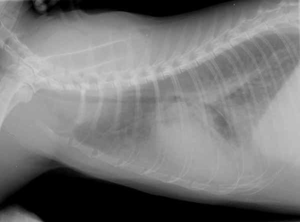
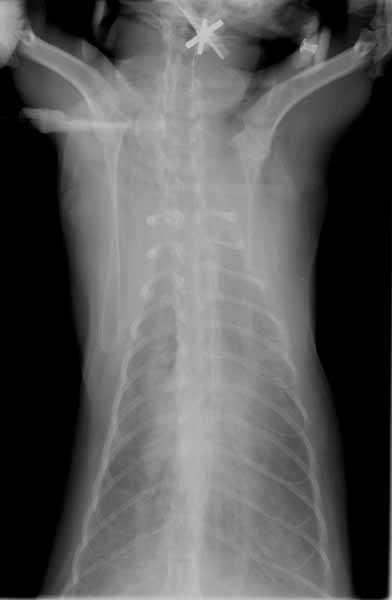
1. The cardiac silhouette is difficult to visualize on the DV view. What is the most likely reason?
2. What type of pulmonary pattern is present on the lateral radiograph?
3. Is this pattern compatible with cardiogenic pulmonary edema in a cat?