Skip to main content
SACM
Home
Cases
ECG Atlas
Case 13 — Echocardiograms
Right Parasternal Long-Axis View
Long-axis view. Electronic calipers measure the RV free wall thickness at 15 mm. RV; LV; LA; RA.
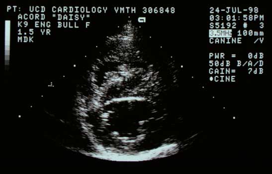
Right Parasternal Short-Axis View at the Ventricles
Short-axis view at the ventricles.
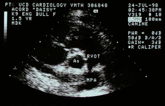
Right Parasternal Short-Axis View at the Base
Short-axis base view. Electronic calipers measure the small pulmonic valve annulus. RVOT; Ao; MPA.
Quiz
1. The findings on this page are characteristic of what congenital abnormality?
Tricuspid valve dysplasia with pulmonary hypertension
Double chambered right ventricle
Pulmonary hypertension due to a right-to-left shunting PDA
Pulmonic stenosis
Check Answer
← ECG
Next: Color Flow →
↑