Case 13 — Coronary Artery Angiograms

Aortic Root Injection
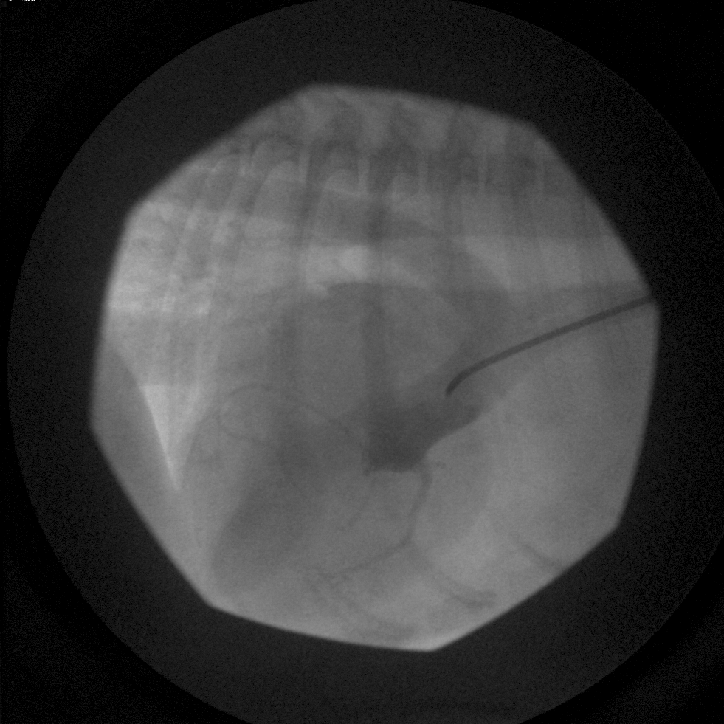
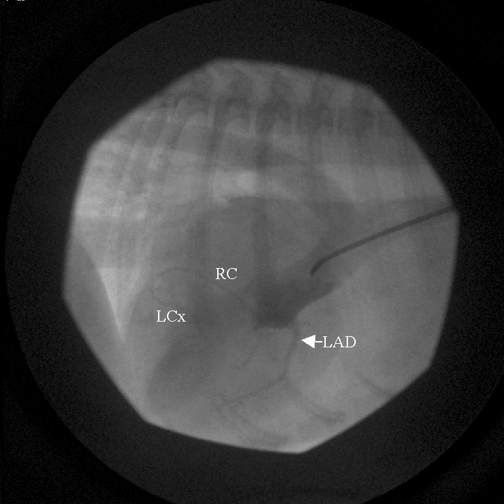
Aortic root angiogram showing contrast injected into the proximal ascending aorta. Only one coronary ostium is visible with all coronary branches originating from it.
Aortic root injection (unlabeled).
Labeled version of the aortic root angiogram. The left anterior descending (LAD) coronary artery is the largest, descending down the front of the heart. The right coronary (RC) artery is dorsal and normal. The left circumflex (LCx) coronary artery is fed by a small branch that courses ventrally then horizontally.
Labeled. LAD (largest, descends anteriorly); RC (dorsal, normal); LCx (fed by small branch coursing ventrally then horizontally).

Selective Injection into the Right Coronary Ostium
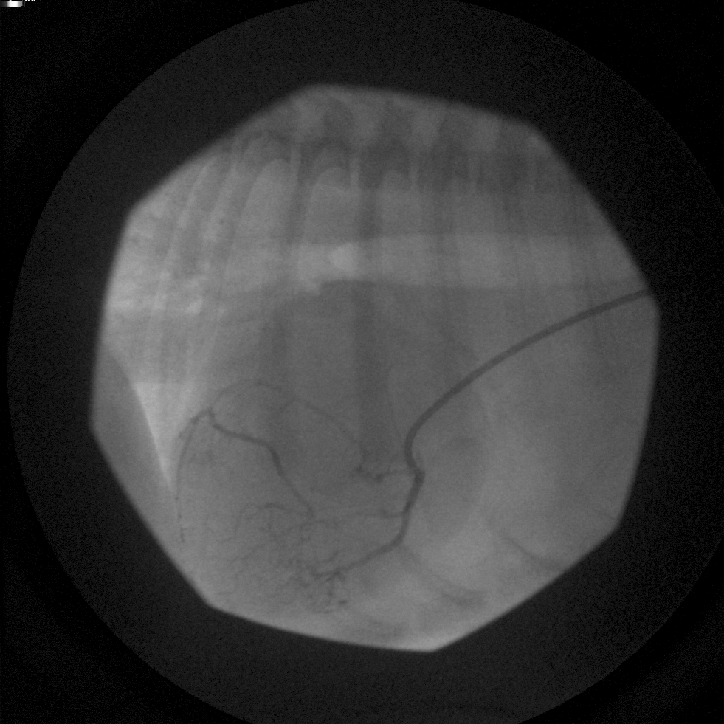
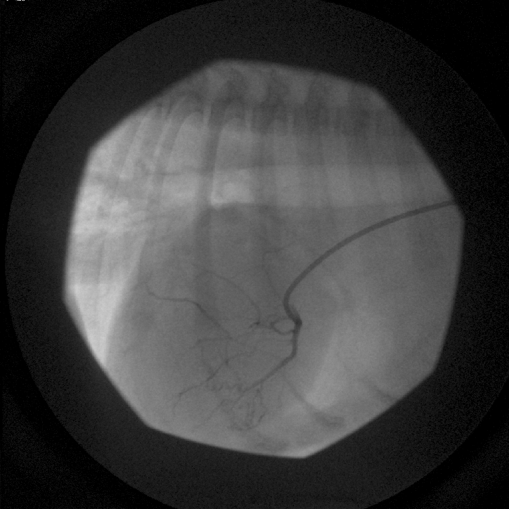
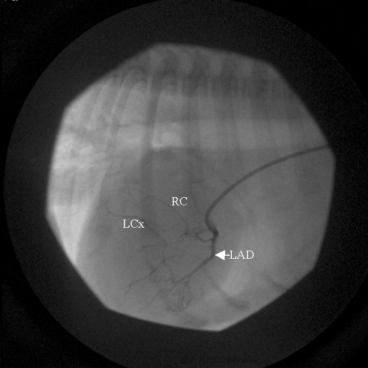
Selective angiogram with catheter in the right coronary ostium. All coronary branches fill simultaneously. The LAD originates from the right ostium. The LCx is fed by a small branch from the LAD and collateral vessels. The upper small branch appears to wrap around the pulmonic valve region.
Selective right coronary ostium injection (unlabeled).
Labeled version showing LAD, RC, and LCx coronary arteries all originating from the right coronary ostium. The LCx feeder branch wraps around the pulmonic valve region.
Labeled. All branches from the right ostium. The LCx feeder wraps around the pulmonic valve region.
Additional angiographic frame taken in systole. The LCx feeder branch has moved dorsally, creating an appearance of encircling the pulmonic valve region.
Systolic frame (unlabeled).
Labeled systolic frame. In systole, the LCx feeder has moved dorsally creating an illusion of connecting to the RC artery and encircling the pulmonic valve region. LAD — left anterior descending.
Labeled. In systole, the LCx feeder moves dorsally, creating an illusion of encircling the pulmonic valve region.

Video Clip of Aortic Root Angiogram

Contrast agent was injected into the root of the aorta. Only one coronary ostium leading to a large coronary artery can be seen on the ventral border of the aortic root. The large artery is the left anterior descending coronary artery. Two smaller arteries can be seen coursing backward. The upper one is the right and the bottom one is the left circumflex.


Video Clip of a Selective Injection into the Right Coronary Ostium
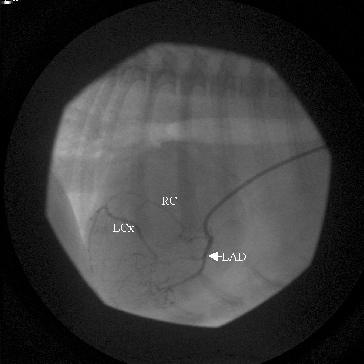
Contrast flows first into the large LAD, then can be seen coursing around the pulmonic valve region to turn into the left circumflex (lower) and right coronary (upper) arteries. At the end, contrast faintly appears in the great cardiac vein immediately above the right coronary artery.