Skip to main content
SACM
Home
Cases
ECG Atlas
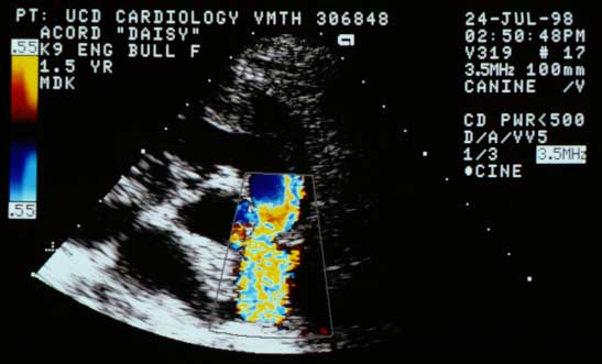
Case 13 — Color Flow Doppler Echocardiograms
Right Parasternal Short-Axis View at the Base
Short-axis base view with color flow Doppler (systole). Turbulent high-velocity flow is visible crossing the stenotic pulmonic valve into the pulmonary artery.
Video of a color flow Doppler echocardiogram from another dog with the same abnormality
Your browser does not support the video tag.
Quiz
1. What does this color flow Doppler picture show?
Aliased flow into the main pulmonary artery
Pulmonic insufficiency
Turbulent, high-velocity flow across a stenotic pulmonic valve
Laminar flow
Check Answer
← 2D Echo
Next: Doppler →
↑