Case 14 — Artificial Pacemaker Implantation

Following placement of the temporary pacemaker, Houdi was anesthetized and a permanent artificial pacemaker was implanted using a transvenous approach via the jugular vein. All pacemaker generators and leads are donated by Medtronic. They donate generators that have gone off “shelf-life” (too long on the shelf for FDA-approved human use). Because they are donated, the cost of implantation is approximately $2,500.

Animated diagram showing pacemaker lead placement through a vein into the right ventricle of a human heart. The lead in Houdi was placed through the jugular vein.
Schematic of pacemaker lead placement. Houdi’s lead was placed through the jugular vein.
Fluoroscopic image showing two leads. The ventral lead is the temporary pacing lead with its tip in the RV apex. The dorsal lead (permanent) has been advanced into the caudal vena cava and needs to be retracted and redirected toward the tricuspid valve.
Temporary lead (ventral) in the RV apex. Permanent lead (dorsal) has been advanced into the caudal vena cava and needs redirecting.
Fluoroscopic image showing the permanent lead withdrawn and readvanced toward the tricuspid valve. The tip is lodged against the valve and bending as force is applied.
Permanent lead redirected toward the tricuspid valve. The tip is lodged against the valve, bending with applied force.
Fluoroscopic image showing the permanent lead has crossed the tricuspid valve with the tip lying in the RV apex alongside the temporary lead.
Permanent lead has crossed the tricuspid valve. Tip in the RV apex alongside the temporary lead.
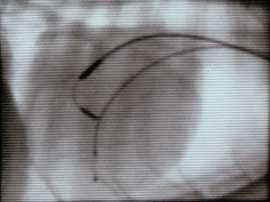
Fluoroscopic image showing the permanent lead in good final position. The tip is angled caudally with enough force to keep it bent against the myocardium. Plastic tines on the tip entwine in the trabeculae. The temporary lead has been withdrawn.
Final position. The tip is angled caudally with tines entwining the trabeculae. Temporary lead has been withdrawn.
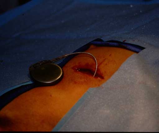
Photograph from another dog showing the pacemaker generator attached to the lead. The lead has been tunneled subcutaneously from the neck (jugular vein) back to the lateral thorax. A skin incision and pocket beneath the cutaneous trunci muscle hold the generator.
Pacemaker generator (from another dog). The lead is tunneled subcutaneously from the jugular vein to a pocket beneath the cutaneous trunci muscle on the lateral thorax.