Case 19 — Doppler Echocardiograms

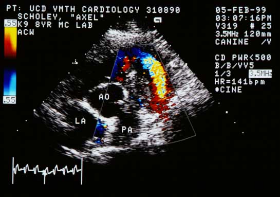
Color flow Doppler showing a pulmonary artery below the aorta (AO), almost totally occluded by the structure. Flow around the structure is markedly reduced and turbulent.
Color flow Doppler. The PA is almost totally occluded by the structure. Residual flow is reduced and turbulent.
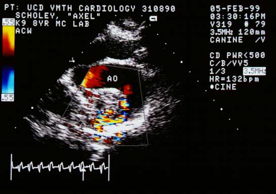
Color flow Doppler in systole showing a larger turbulent flow pattern in the right ventricular outflow tract, probably large enough to explain the heart murmur heard on physical examination.
Color flow Doppler (systole). Turbulent flow in the RVOT, probably explaining the auscultated murmur.
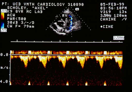
Continuous wave Doppler measuring peak systolic flow velocity in the right ventricular outflow tract at approximately 2 meters per second.
CW Doppler. Peak systolic velocity in the RVOT: ~2 m/s.
Quiz

1. What is the pressure gradient across the mass from the right ventricle to the pulmonary artery?