Case 19 — Two-Dimensional Echocardiograms

Right Parasternal Cross-Sectional View at the Base

Right parasternal cross-sectional basilar view showing the aorta (AO), left atrium (LA), and right ventricle (RV). An arrow points to a structure in the right ventricular outflow tract.
Basilar view. AO; LA; RV. Arrow: structure in the right ventricular outflow tract.

Right Parasternal Cross-Sectional View at the Ventricles

Cross-sectional view at the ventricular level. RV and LV labeled. Arrows point to the ventral border of the same structure seen in the basilar view.
Ventricular level. RV; LV. Arrows: ventral border of the same structure.

Right Parasternal Cross-Sectional View at the PA Branches

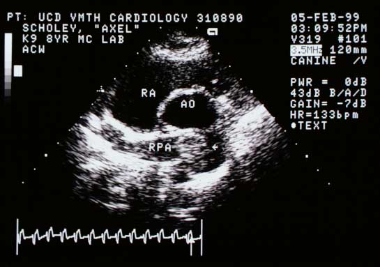
Cross-sectional view at the pulmonary artery branches showing the top of the structure in the main pulmonary artery, at least partially occluding the right caudal lobar pulmonary artery branch (RPA). AO — aorta; RA — right atrium.
PA branch level. The structure extends into the main PA and partially occludes the RPA. AO; RA.

Video of an echocardiogram from a different dog with the same abnormality


Quiz

1. Choose the two primary rule-outs for the structure in the RVOT and proximal main pulmonary artery.