Skip to main content
SACM
Home
Cases
ECG Atlas
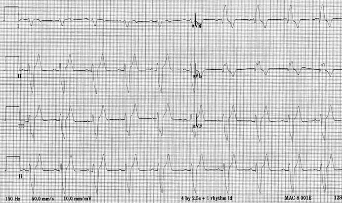
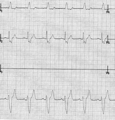
Case 19 — ECGs
Six limb leads (I, II, III left; aVR, aVL, aVF right) with lead II continuous along the bottom.
Chest leads V
1
and V
2
with lead II at bottom.
Quiz
1. What conduction abnormality is present?
Right bundle branch block
Left bundle branch block
Second degree AV block
First degree AV block
Check Answer
← Overview
Next: 2D Echo →
↑