Case 4 — Electrocardiogram (ECG)

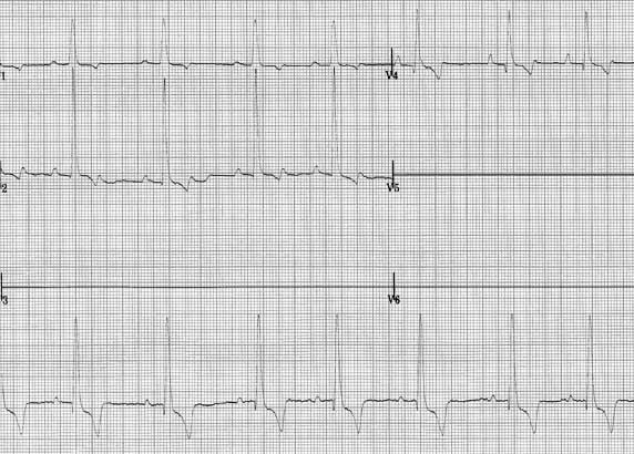
This ECG provides the six limb leads plus three chest leads (V1, V2, and V4). Calibration markers are to the left on the limb leads and show that 1 cm = 1 mV. Paper speed is 50 mm/second.
This ECG provides the six limb leads plus three chest leads (V1, V2, and V4). Calibration markers are to the left on the limb leads and show that 1 cm = 1 mV. Paper speed is 50 mm/second.