Case 4 — Echocardiograms of the Left Ventricle
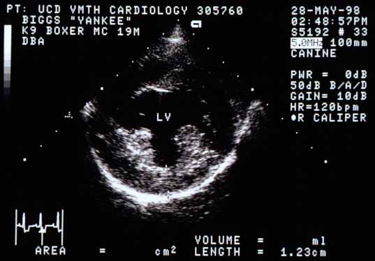
This is a cross-sectional view of the left ventricle taken from a right parasternal position. The left ventricular free wall is 1.23 cm thick (normal for this sized dog is 9 to 10 mm). The papillary muscles, especially the caudal one (to the left on the screen), are hyperechoic, probably because there is increased fibrous (scar) tissue in them.

This right parasternal longitudinal four-chamber view shows normal left ventricular (LV), left atrial (LA), and right atrial (RA) chamber sizes. The thickness of the interventricular septum (cursors) is increased to 1.36 cm.