There was no evidence of pleural effusion or pulmonary edema on post mortem examination,
confirming that Alice did not die of heart failure and so most likely died suddenly.
Alice’s heart weighed 31 grams. The normal heart weight for
an adult cat should be less than 20 grams. Cats with severe hypertrophic cardiomyopathy have
a heart weight greater than 30 grams. The worst heart from a cat with severe HCM that I have
seen weighed 38 grams.
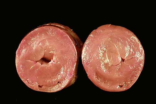
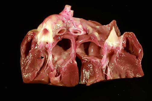
Unopened heart viewed from the front. The left auricle (top right) is enlarged compared to the right auricle (left).Cross-sectional view of the left ventricle. The heart is in rigor (post-mortem contracture), so wall thicknesses are comparable to a systolic echocardiographic frame. Note the near-obliteration of the LV cavity and the extremely enlarged papillary muscles.Longitudinal section through the left atrium, mitral valve, and aorta. The arrow points to the narrowed left ventricular outflow tract (LVOT) between the basilar interventricular septum and the anterior mitral valve leaflet. The enlarged left atrium lies above the mitral valve.