Case 7 — Echocardiograms
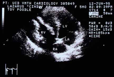
Four-chamber view (left apical position). The right ventricular free wall (left of RV label) is extremely thickened. The subendocardium is severely hyperechoic, suggesting damage and fibrosis. RV — right ventricle; LV — left ventricle; RA — right atrium; LA — left atrium.
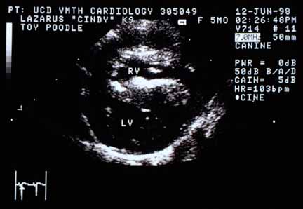
Cross-sectional view (right parasternal position). Again shows the severely thickened right ventricular free wall and the hyperechoic subendocardium.
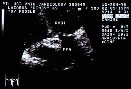
View of the pulmonic valve (between RVOT and MPA), taken in systole. The valve cusps are thicker than normal and immobile — they have not opened.
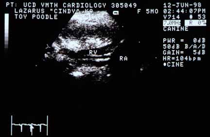
View of the tricuspid valve showing long and thickened chordae tendineae (immediately below the RV label). RA — right atrium.
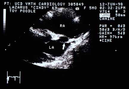
View of the atrial septum (right parasternal). Two walls of the atrial septum are visible, separated by a hole (arrow).
Video of an echocardiogram from another dog with the same disease
Close-up of the pulmonary valve.