Case 8 — Color Flow Doppler Echocardiography

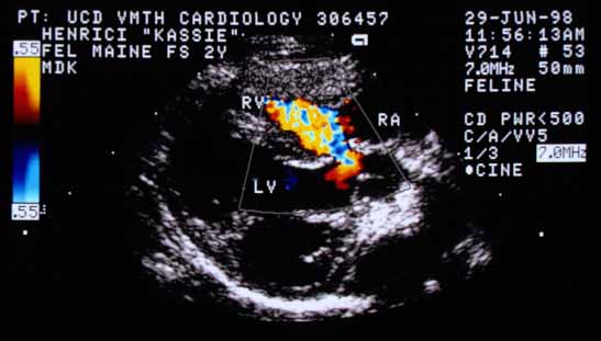
Color flow Doppler image recorded in systole from a long-axis view. Flow starts in the left ventricle (LV) beneath the aorta as laminar flow toward the transducer (homogeneous red). Just before the upper interventricular septum, the color turns gold as velocity nears 0.55 m/s (Nyquist limit), then aliases to blue. As it passes through the defect, it becomes a mosaic of colors and projects into the right ventricle (RV). RA — right atrium.
Color flow Doppler (systole, long-axis). Laminar flow (red) in the LV aliases to gold/blue at the upper septum, then becomes a mosaic turbulent jet projecting through the defect into the RV. RA — right atrium.
Color flow Doppler in cross-sectional view. Flow starts beneath the aorta (Ao) in the left ventricle as red laminar flow, then turns into a high-velocity mosaic jet that strikes the right ventricular free wall and turns to the right into the right ventricular outflow tract, where it loses momentum and becomes blue laminar flow away from the transducer.
Color flow Doppler (cross-section). Flow beneath the Ao begins as laminar (red), becomes a mosaic jet through the defect striking the RV free wall, then turns into blue laminar flow in the RVOT.

Video of an echocardiogram from a different cat with the same abnormality
Same video at half speed


Quiz

1. Is the direction of this jet compatible with the location of this cat’s heart murmur?


↑