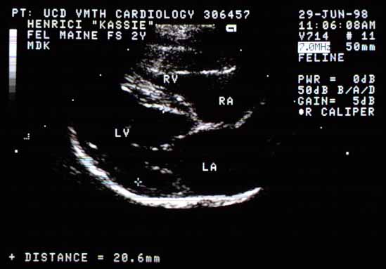
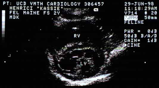
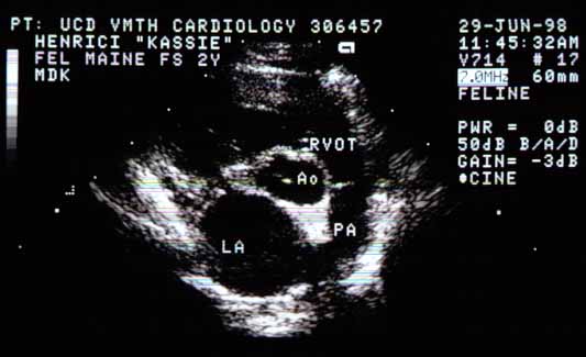
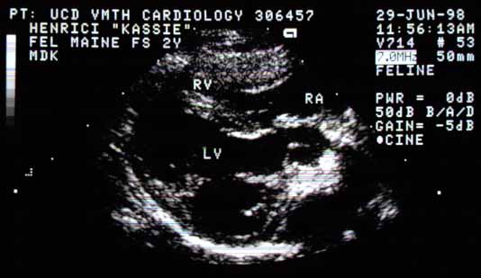
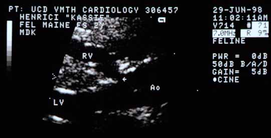
Right parasternal long-axis view (diastole). LV diastolic diameter is increased to 21 mm (normal ≤17 mm). The RV free wall is severely thickened. LA and RA are enlarged.Right parasternal cross-sectional view. The RV chamber is enlarged and the RV free wall thickened. Abnormal chordal structures emanate from the papillary muscles in the LV chamber.Cross-sectional base view (diastole). LA is moderately enlarged relative to Ao. The pulmonic valve (between RVOT and PA) is closed.Long-axis view of LV and aorta. A region of discontinuity is visible between the upper interventricular septum and the base of the aorta.Close-up of the base of the interventricular septum. A defect (arrow) is visible between the base of the septum and the aortic root.
Video of an echocardiogram from a different cat with the same abnormality