Case 8 — M-Mode

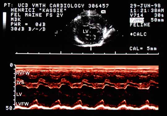
M-mode echocardiogram showing an enlarged left ventricular (LV) cavity of approximately 21 mm in diastole. The left ventricular free wall (LVFW) motion is vigorous but end-systolic diameter is increased to approximately 12 mm, suggesting decreased contractility. The right ventricular free wall (RVFW) is severely thickened. The interventricular septum (IVS) moves abnormally, flattening in systole and becoming round again in diastole.
M-mode echocardiogram. LV diastolic diameter ~21 mm (enlarged); end-systolic diameter ~12 mm (increased, suggesting myocardial failure). RVFW is severely thickened. The IVS flattens in systole and becomes round in diastole.

The left ventricular free wall (LVFW) motion is vigorous, yet the increased end-systolic diameter is probably due to decreased left ventricular contractility (myocardial failure). When examined on the two-dimensional echocardiogram, the abnormal septal motion was appreciated as the interventricular septum flattening in systole and becoming round again in diastole.


Quiz

1. What is the flattening of the interventricular septum in systole due to?


↑