Case 9 — Figures

Normal Mitral Valve

Diagram of normal mitral valve anatomy showing the leaflets, annulus, chordae tendineae, and papillary muscles.
Normal mitral valve anatomy.

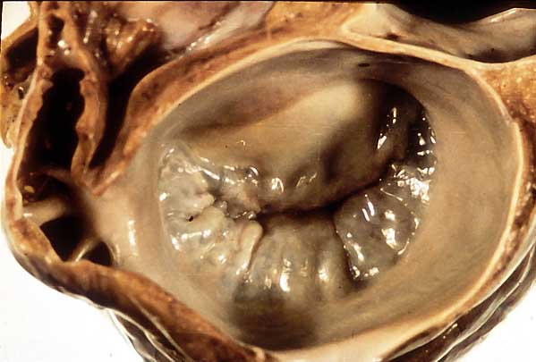
Pathology Specimen of Severe Myxomatous Mitral Valve Disease

Gross pathology specimen showing a mitral valve with severe myxomatous degeneration. The leaflets are markedly thickened and nodular.
Pathology specimen of severe myxomatous mitral valve disease.

Normal Mitral Valve Histology
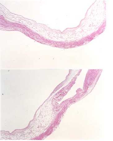
Histological section of a normal mitral valve showing the atrialis, spongiosa, and fibrosa layers.
Normal mitral valve histology.

Histopathology of Severe Myxomatous Mitral Valve Disease
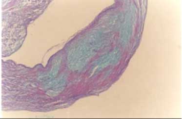
Histological section showing severe myxomatous degeneration of the mitral valve with expanded spongiosa layer and degenerated fibrosa.
Histopathology of severe myxomatous mitral valve disease.

Schematic Drawing of Moderate Mitral Regurgitation
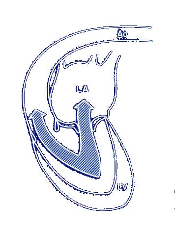
Schematic diagram showing the hemodynamics of moderate mitral regurgitation with blood leaking from the left ventricle back into the left atrium during systole.
Schematic of moderate mitral regurgitation.

Contrast Injection (Angiogram) into the Left Ventricle of a Dog with Severe Mitral Regurgitation
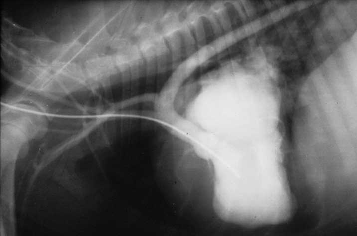
Left ventricular angiogram from a dog with severe mitral regurgitation showing contrast filling both the left ventricle and the left atrium during systole.
Left ventricular angiogram showing severe mitral regurgitation.

Color Flow Doppler Echocardiogram from a Dog with Severe Mitral Regurgitation
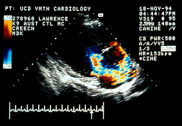
Color flow Doppler echocardiogram showing a large turbulent jet of mitral regurgitation projecting from the mitral valve into the left atrium.
Color flow Doppler showing severe mitral regurgitation.

↑