Case 13 — Case Discussion
This dog had severe pulmonic stenosis. The chest radiographs were not very helpful, primarily because of the bulldog chest configuration. Bulldog chests are always difficult to interpret.
The ECG was typical of severe right ventricular enlargement, with deep S waves in leads I, II, aVF, and V4. The P waves in lead II were too wide (0.05 seconds) and notched — typically seen with left atrial enlargement, but this dog did not have LA enlargement, making this a false positive finding. The P-wave widening may be explained by disruption of the intraatrial conduction system.
The echocardiogram showed the pulmonic stenosis had at least an annular component (markedly reduced annulus size) and a valvular component.
In many English bulldogs, pulmonic stenosis is of the subvalvular type and is either associated with or caused by an R2A coronary artery anomaly.
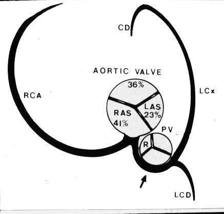
In Daisy, the coronary ostium appeared similarly placed but the left coronary artery appeared to course between the pulmonary artery and the aorta. Previous experience with balloon valvuloplasty in English bulldogs with R2A anomalies resulted in sudden death from avulsion of the left coronary artery. Even if the artery passed between the PA and aorta, balloon inflation could cause cessation of coronary flow. Consequently, balloon valvuloplasty was not performed.
The owner’s primary complaint was the syncopal events. Differential diagnoses included tachyarrhythmia, bradyarrhythmia, and vasodilation. The event recorder showed no arrhythmia during a syncopal episode, ruling out an arrhythmia. It was assumed Daisy was having vasodepressor syncope (acute vasodilation causing severe hypotension). However, her events were not consistently precipitated by exercise or excitement, and the observation that some episodes were preceded by “gagging” suggested an additional possibility — stimulation of vagal afferents in the pharynx.
Follow-Up
No treatment was initially prescribed. About 6 weeks after hospitalization, Daisy had only two more syncopal events. The first occurred when she was outside on a hill and she literally rolled over 3–4 times down to the deck. She recovered quickly. The last event was more severe — she fell over while the owner was talking to his neighbor, was white, eyes open but unresponsive, and urinated and defecated. The event lasted an estimated 3–5 minutes (much longer than previous episodes). Daisy did not bounce right back and took a couple of days to seem right.
The observation that some episodes were preceded by a “gagging” sound led to a surgical evaluation. Daisy was examined by the soft tissue surgeons and found to have an elongated soft palate, stenotic nares, and everted saccules. She underwent surgery to correct these abnormalities. At the time of the last update (four months following the procedure), she was doing very well and had not had another syncopal event.