Skip to main content
Case 17 — Color Flow Doppler
2D Echocardiogram (Left-Sided View)
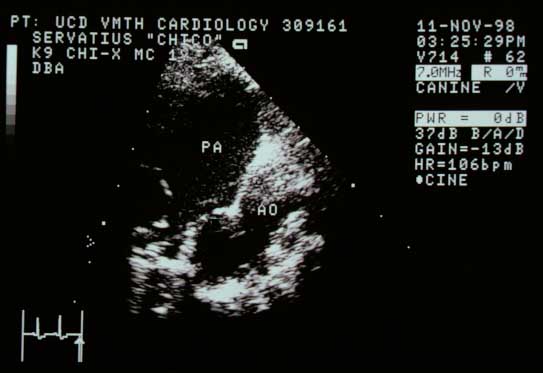 Left-sided view. Proximal descending aorta (AO) and main pulmonary artery (PA) with a structure with an opening between them.
Color Flow Doppler (Same View)
 Color flow Doppler (systole). Continuous flow from aorta through the ductus into the PA, with turbulent jet extending to the pulmonic valve.
Color flow Doppler from a different dog with the same abnormality
Same video at half speed
↑