Case 17 — Two-Dimensional Echocardiograms

Right Parasternal Long-Axis View

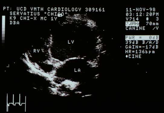
Right parasternal long-axis view showing subjectively enlarged left ventricular (LV) chamber. The left atrium (LA) is similarly enlarged. The right ventricular (RV) chamber is normal.
Long-axis view. LV chamber subjectively enlarged. LA similarly enlarged. RV normal.

Left Apical Four-Chamber View

Left apical four-chamber view showing the same findings as the long-axis view — enlarged left heart chambers.
Left apical four-chamber view. Same findings as above.

Right parasternal short-axis view of the left ventricle from another dog with the same abnormality

The LV chamber is much larger than normal in diastole with similarly enlarged end-systolic diameter, indicating myocardial failure despite normal shortening fraction.

Right Parasternal Cross-Sectional Basilar View

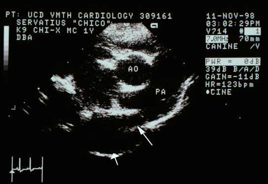
Right parasternal cross-sectional basilar view showing the main pulmonary artery (PA) wider than normal compared to the aorta (AO).
Basilar view. The main pulmonary artery (PA) is wider than normal relative to the aorta (AO).
Same basilar view with arrows. Short fat arrow points to the descending aorta. Longer arrow points to a structure between the aorta and pulmonary artery — the patent ductus arteriosus.
Same view. Short arrow: descending aorta. Long arrow: structure between the aorta and PA.

Quiz

1. What is the structure being pointed to in the last image?

2. The left ventricular chamber is enlarged and the wall thickness is normal. How would you characterize this ventricle?