Skip to main content
SACM
Home
Cases
ECG Atlas
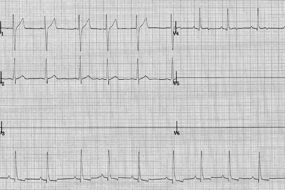
Case 17 — Electrocardiogram (ECG)
Six limb leads (50 mm/s, 5 mm = 1 mV). R wave in lead II ~4 mV (normal 1–2.5 mV).
Chest leads: V
1
(top left), V
2
(below), V
4
(top right).
Quiz
1. There is evidence of one chamber being enlarged. Which one?
Right atrium
Right ventricle
Left atrium
Left ventricle
Check Answer
← Radiographs
Next: 2D Echo →
↑