Case 8 — Case Discussion
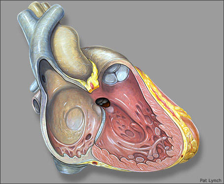
Unfortunately Kassie was given a relatively poor prognosis. Her measured systemic systolic arterial blood pressure using a Park’s Doppler unit was 105 mmHg. Since the pressure gradient across her ventricular septal defect (VSD) was only 45 mmHg, this meant that her right ventricular systolic pressure was approximately 60 mmHg. Since she had no evidence of pulmonic stenosis, this meant that her pulmonary artery systolic pressure was also approximately 60 mmHg. Normal systolic pulmonary artery and right ventricular pressures are around 15 to 20 mmHg. This means that Kassie had pulmonary hypertension, graded at moderate to severe.
An increase in systolic right ventricular and pulmonary artery pressure in a VSD can occur either because of an increase in flow or an increase in resistance (or more appropriately impedance). To increase pressure to the degree seen in Kassie, flow would have to be increased markedly (probably more than three times normal). The defect in Kassie’s interventricular septum was not large, so this possibility seemed less likely. However, the increased diastolic diameter of the left ventricular chamber suggested that the flow and the defect were larger than was suggested by the defect size. Consequently, increased flow could have been contributing to the increased systolic pressure. However, the pulmonary hypertension was almost assuredly also due to pulmonary vascular disease. Pulmonary vascular disease occurs secondary to increased pulmonary blood flow in patients with a VSD. Although this usually requires large flows, occasionally it can occur with more modest flows.
Treatment of a large VSD in a cat is generally medical or surgical palliation. Surgical closure of a VSD requires cardiopulmonary bypass and open heart surgery — neither of which are technically feasible in a domestic cat. Medical therapy is often aimed at treating signs of left heart failure. However, an arteriolar dilator such as hydralazine can also be used. The amount of left-to-right shunting across a VSD depends on the size of the defect and the relative resistances (impedances) of the pulmonary and systemic circulations. If the systemic vascular resistance is decreased with hydralazine, more blood flows into the systemic circulation and less flows across the VSD into the pulmonary circulation. Alternatively, a band can be placed around the main pulmonary artery at surgery to create supravalvular pulmonic stenosis, which increases pulmonary vascular resistance and decreases shunt flow. Neither a pulmonary artery band nor hydralazine were considered viable options for treating Kassie — both were more likely to create right-to-left flow than to improve hemodynamics. Consequently, she was sent home with no therapy but with instructions to reexamine her in six months to monitor disease progression. Most likely her pulmonary disease will get worse with time and will ultimately result in right-to-left shunting, systemic hypoxemia, and eventually polycythemia that may require phlebotomy for control.
The latest follow-up was 3.5 months after the initial visit. At that time, the owner reported that Kassie was doing very well and showing no clinical signs whatsoever. She was reminded to have the PCV rechecked within the first 6 months and to report back if there were any problems.