Case 9 — Color Flow Doppler
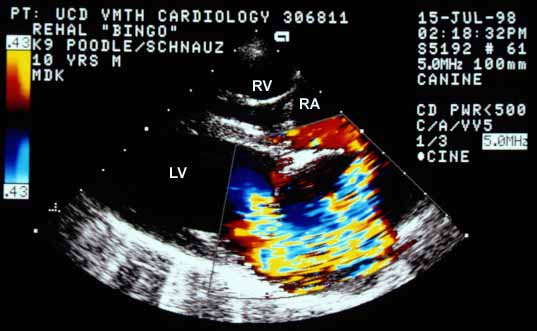
Color flow Doppler in systole from a right parasternal long-axis view. Blood flow starts as laminar flow away from the transducer (blue) in the left ventricle (LV), aliases to gold near the mitral valve, then becomes a mosaic high-velocity jet projecting back into the left atrium (LA). The jet is eccentric, striking the left lateral wall of the LA and wrapping around within the chamber. RV — right ventricle; RA — right atrium.
Systolic color flow Doppler (right parasternal long-axis). Laminar flow in the LV aliases at the mitral valve, becoming an eccentric mosaic jet that strikes the LA wall and wraps around the chamber. RV; RA.
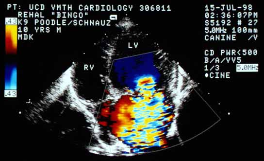
Color flow Doppler in systole from a left apical four-chamber view. Flow in the LV progresses from blue (laminar) through lighter blue, gold, red, and back to blue as velocity increases toward the Nyquist limit (0.43 m/s). The flow narrows at the mitral valve defect forming the jet base, which is quite wide. The jet is large, filling the entire left atrium. RV — right ventricle.
Systolic color flow Doppler (left apical four-chamber). Flow accelerates through the color spectrum toward the Nyquist limit (0.43 m/s). The wide jet base at the mitral valve defect and the large jet filling the entire LA indicate severe disease. RV.
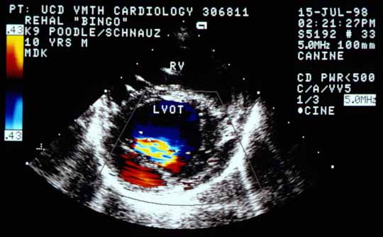
Right parasternal cross-sectional view of the mitral valve region showing the base of the color flow jet as it crosses through the mitral valve defect, below the left ventricular outflow tract (LVOT). The jet base appears very large. RV — right ventricle.
Cross-sectional view of the mitral valve region showing the large base of the color flow jet crossing through the mitral valve defect (below the LVOT). RV.

Video clip of a color flow Doppler echocardiogram from another dog with the same abnormality

Note the color flow jet that wraps around within the left atrium, first hugging the lateral wall and then coursing along the top of the left atrium.


Quiz

1. The color flow Doppler pattern seen on this page is diagnostic of what functional cardiac abnormality?

2. Based on the size of this dog’s left atrium, the size of the orifice in the mitral valve, and possibly the size of the color flow jet, how would you classify the severity?


↑