Case 9 — M-Mode Echocardiograms

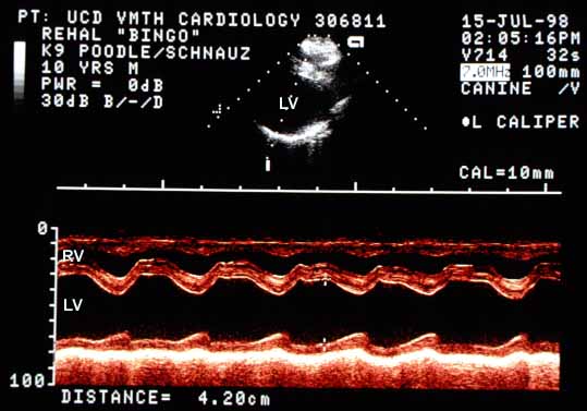
M-mode echocardiogram of the left ventricle. End-diastolic diameter (EDD) is 4.2 cm (normal ~3.0 cm). End-systolic diameter (ESD) is 2.4 cm (normal ~2.0 cm). Wall excursion is 1.8 cm, increased for this size dog. The interventricular septum excursion is greater than normal. Free wall motion appears normal. Wall thicknesses (IVS and LVFW) are normal at 8 mm.
M-mode of the left ventricle. EDD = 4.2 cm (normal ~3.0 cm); ESD = 2.4 cm (normal ~2.0 cm). IVS excursion is increased. Wall thicknesses are normal (8 mm).
M-mode echocardiogram taken with the beam crossing the mitral valve, showing the labeled excursion points A through F. The E-point to septal separation (EPSS) is mildly increased to 6 mm (normal ~3 to 4 mm for this size dog).
M-mode of the mitral valve with excursion points labeled (A–F). The E-point to septal separation (EPSS) is mildly increased to 6 mm (normal ~3–4 mm).
Quiz

1. What is the shortening fraction in this dog?

2. The increase in left ventricular end-diastolic diameter with normal wall thicknesses is indicative of what?

3. The most likely reason for the mild increase in end-systolic diameter is:


↑