Case 9 — Thoracic Radiographs

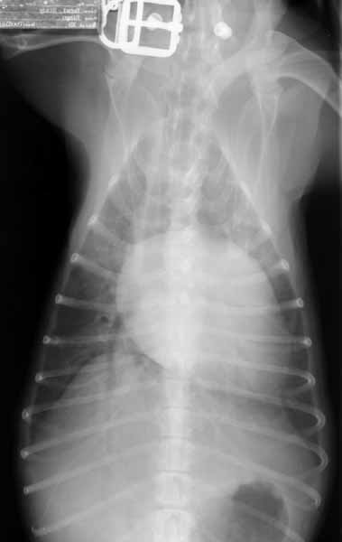
DV

Dorsoventral thoracic radiograph showing severe left atrial enlargement. The left auricle extends beyond the cardiac silhouette at the 2-to-3 o'clock position. The mainstem bronchi are wider apart than normal (bowlegged cowboy sign) due to the enlarged left atrium pushing them apart.
Dorsoventral (DV) thoracic radiograph.

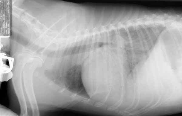
Right Lateral

Right lateral thoracic radiograph showing severe left atrial enlargement (the body of the left atrium touches the spine). A caudodorsal pulmonary infiltrate pattern is present, consistent with cardiogenic pulmonary edema. The cranial lung lobes are spared.
Right lateral thoracic radiograph.

Quiz

1. What cardiac structure is enlarged more than all the others?

2. The pulmonary pattern is consistent with what?

↑