Case 11 — Two-Dimensional Echocardiograms

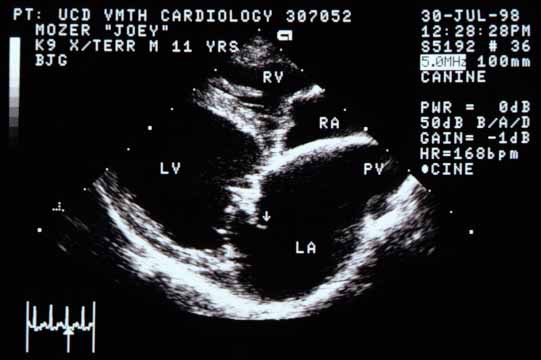
Right parasternal long-axis view of the four chambers. The left ventricular (LV) chamber appears enlarged. The left atrium (LA) is even larger. The right ventricle (RV) and right atrium (RA) may also be enlarged but not to the degree of the left heart. The septal leaflet of the mitral valve is markedly thickened. The pulmonary vein (PV) entering the LA is markedly distended. An arrow points to a structure originating from the mitral valve projecting back into the LA.
Right parasternal long-axis view. LV enlarged; LA even larger. The septal mitral valve leaflet is markedly thickened. The pulmonary vein (PV) is markedly distended. Arrow: structure from the mitral valve projecting into the LA.
Close-up view of the upper left ventricle (LV), lower left atrium (LA), and mitral valve. An arrow points to the same abnormality as above — a structure projecting from the mitral valve into the left atrium.
Close-up of the mitral valve region. Arrow indicates the same structure projecting into the LA. LV; LA.
Left apical four-chamber view showing all four chambers enlarged with left heart size predominating. An arrow points to what appears to be a defect in the atrial septum.
Left apical four-chamber view. All chambers enlarged; left heart predominates. Arrow: apparent defect in the atrial septum.
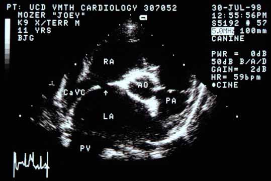
Right parasternal cross-sectional view of the base of the heart showing the markedly enlarged left atrium (LA) compared to the aorta (Ao). The caudal vena cava (CaVC) enters the right atrium (RA). There again appears to be a defect in the interatrial septum.
Cross-sectional base view. The LA is markedly enlarged relative to the Ao. The caudal vena cava (CaVC) enters the RA. A defect in the interatrial septum is again apparent.
Quiz

1. There is a defect in the interatrial septum seen in two views and therefore most likely real. What could cause this defect?

2. What is the arrow pointing to in the top two images?